Life Sciences
An increasing number of biopharmaceutical companies are looking for an AI-imaging partner. AI-powered imaging-based solutions can predict which patients will respond to a drug, find the right candidates for clinical studies, stratify patients with different risks and accelerate drug development programs.
Applications
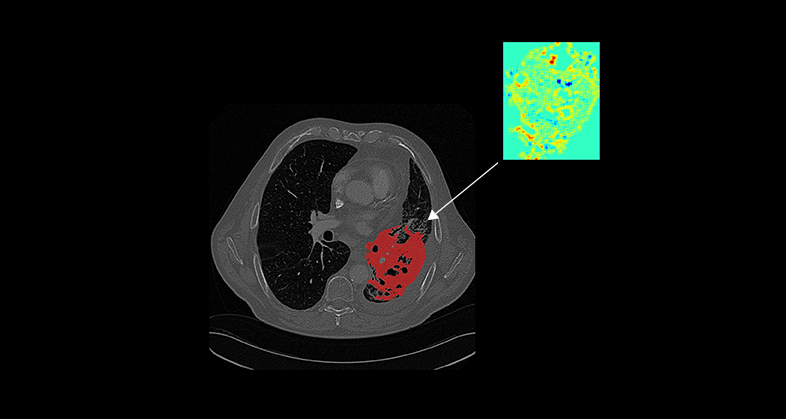
Oncology
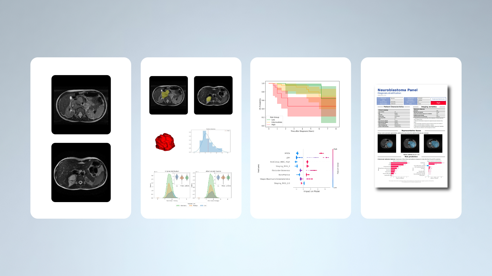
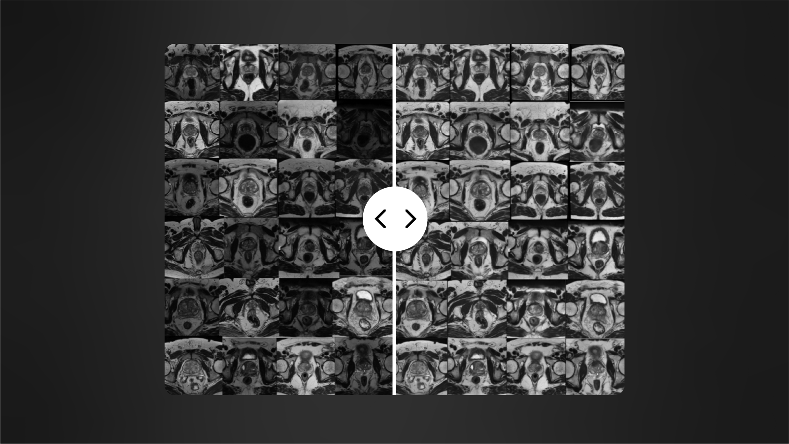
Our tissue-agnostic AI methods are applied at each stage of drug development programs. First we apply our AI techniques to harmonize image quality across hospitals and scanner models, then we perform automated organ and lesion detection. A tissue characterization follows to extract a signature based on the most relevant radiomics-based and deep features. These features are then used to develop predictive models of tumor growth, treatment response, overall survival, disease free survival, among many other patient outcomes.
Immunology
Our AI-based tools provide patients with an early diagnosis for rheumatic diseases in the main imaging modalities. We support the digital transformation of therapy indication either during drug-development phases or in post-approval scenarios, deploying an integrated offering to healthcare providers.
Neuroscience
We apply our AI-based methods for automated segmentation and quantification of brain tissues and abnormal lesions. We assess differences in the volume of brain regions in neurodegenerative and inflammatory diseases and in metabolic disorders, extracting surrogate imaging biomarkers.
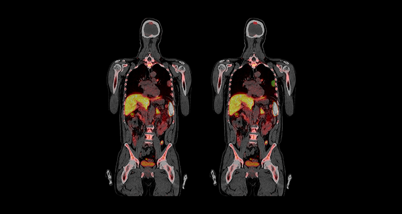
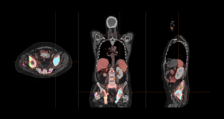
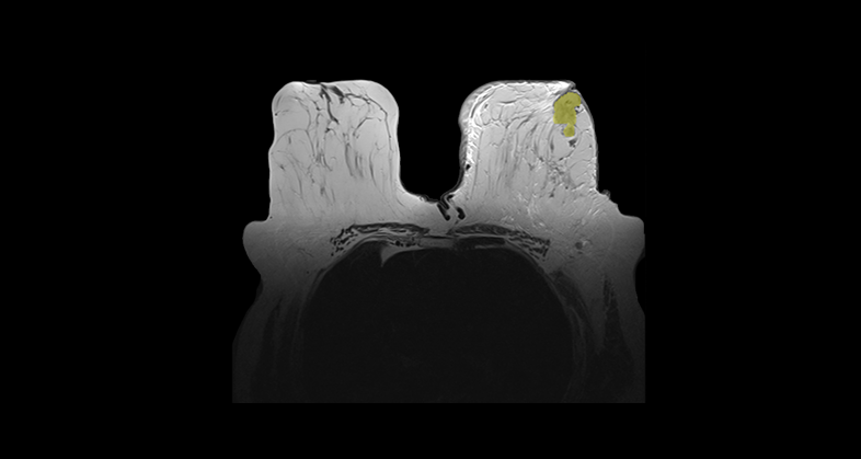
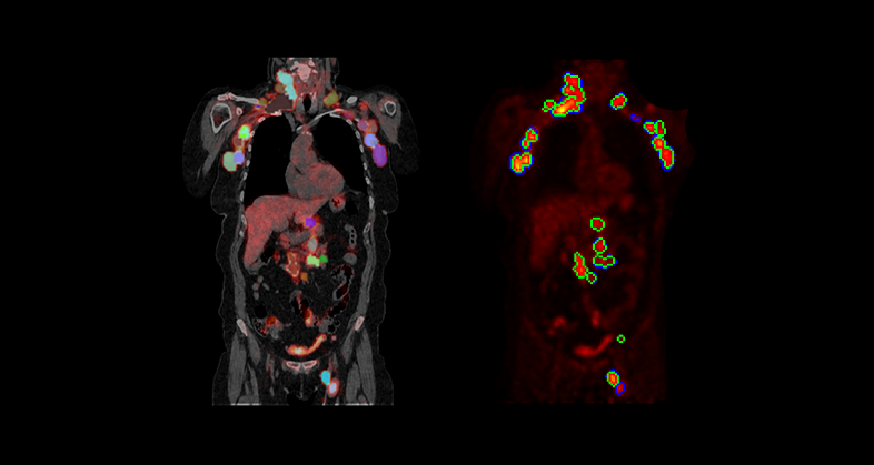
Hematology
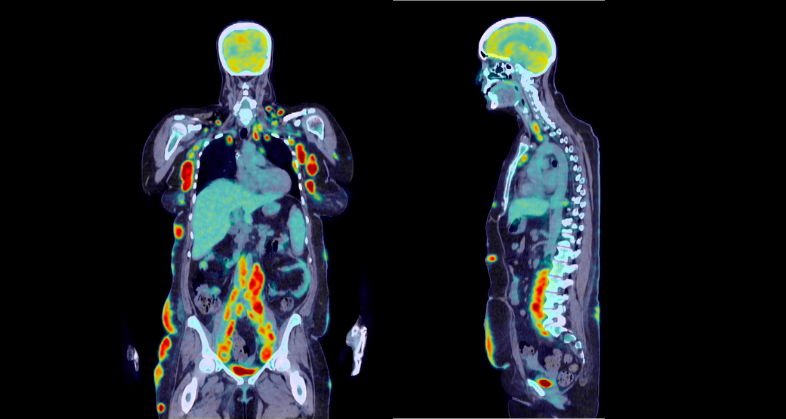
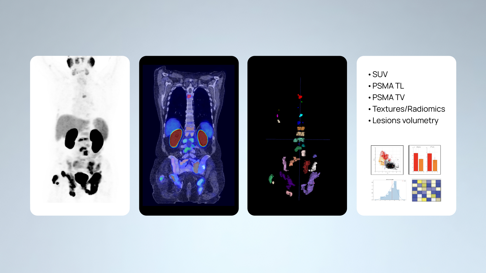
We provide automated segmentation methods for lesions and organs in whole body PET/CT examinations. The most frequent clinical scenarios are lymphoma and multiple myeloma, extracting lesion-level features, which, combined at the patient level, can help in predicting treatment response in standard or advanced therapies as well as other phenomena like cytokine release syndrome (CRS) or Immune effector cell-associated neurotoxicity syndrome (ICANS) in cell therapies such as Chimeric Antigen Receptor T-cell therapies (CAR-T).
Case Studies
-
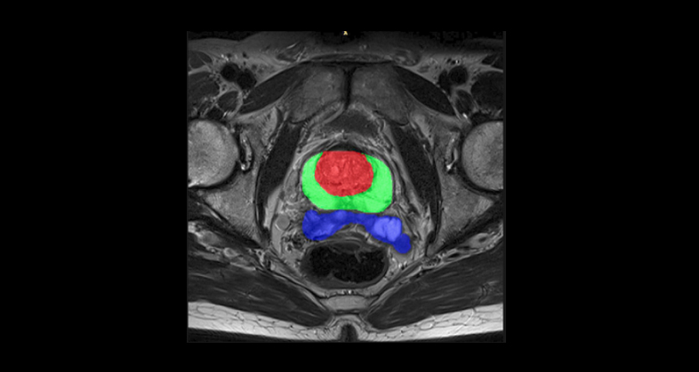
Oncology
Prediction of metastatic relapse in intermediate/high risk localized prostate cancer patients from staging medical images and clinical variables (PROVIDENCE study)
Read more
-
Immunology
Predicting immunotherapy outcomes in patients with advanced NSCLC
Read more
-
Immunology
Predicting immunotherapy response in advanced solid tumors using quantitative imaging features extracted from immune PET scans
Read more
-
Immunology
Oncology
Informing patient stratification strategies for pivotal immunotherapy study of Metastatic Prostate Cancer
Read more
-
Immunology
Empowering neuroblastoma risk stratification through AI algorithms
Read more
-
Immunology
Oncology
Developing a radiomics-based tool for predicting pathological complete response (pCR) to neoadjuvant chemotherapy in breast cancer
Read more
-
Hematology
Oncology
Predicting survival, neurotoxicity, and response in B-Cell lymphoma patients treated with CAR-T therapy using an imaging features-based model
Read more
-
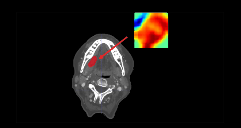
Oncology
CT-based clinical-radiomics model to predict progression and drive clinical applicability in locally advanced head and neck cancer
Read more
-
Hematology
Oncology
Prognostic value of genetic alterations and 18F-FDG PET/CT imaging features in diffuse large B cell lymphoma
Read more

Do you want to be updated with our news?
By pressing submit you accept the privacy policy and the legal notice of the Quibim website.