QP-Brain®

The evolution in brain MRI analysis
With an aging population and the escalating burden of neurodegenerative diseases, there’s a growing need for more accurate assessments to detect early changes in the volume of different brain structures, WMH (white matter hyperintensities) load, and location. QP-Brain® is an AI-powered tool that redefines brain MRI analysis by providing quantitative evaluations for enhanced detection and a better understanding of brain atrophy and lesions.
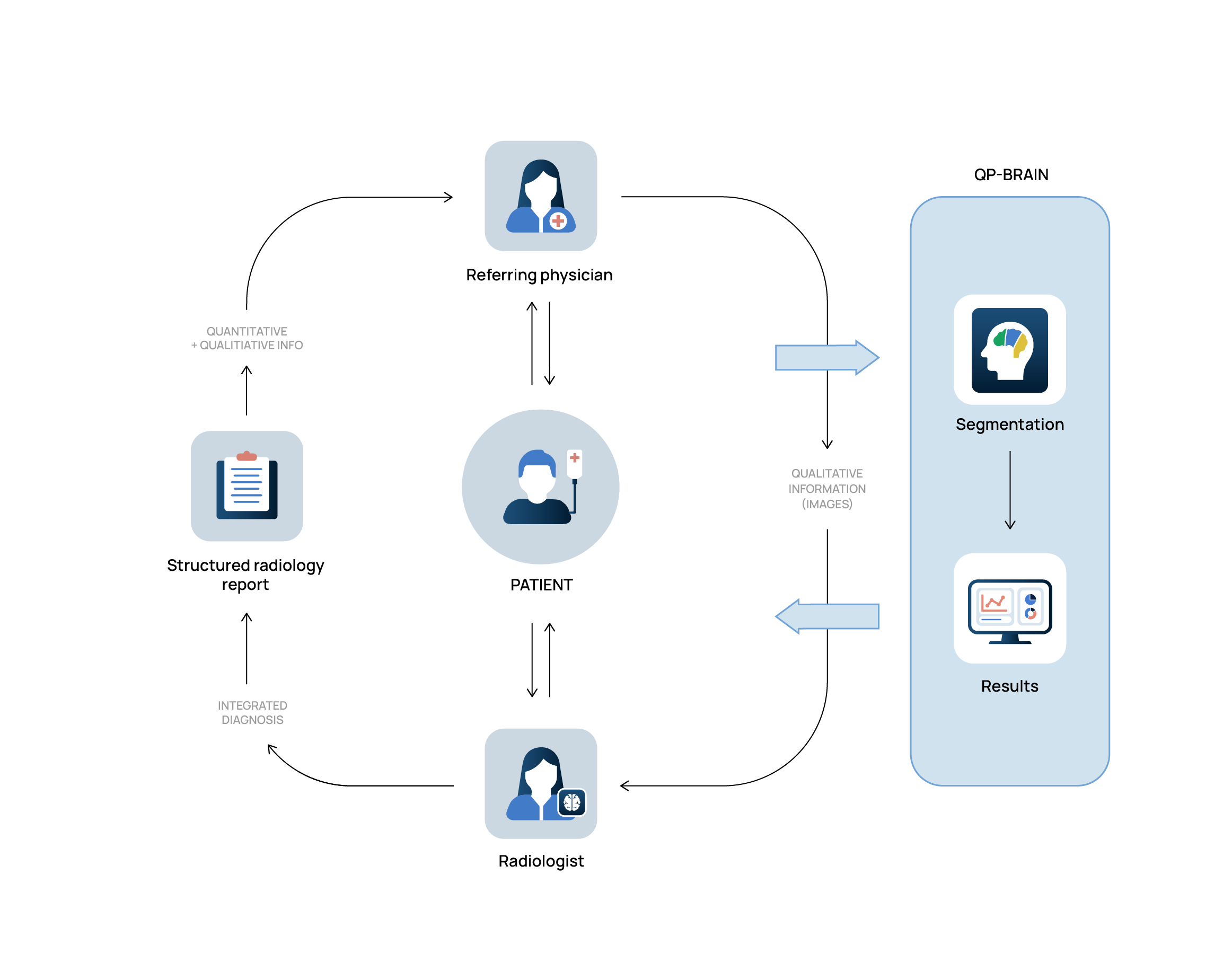
QP-Brain® automates the quantitative analysis of patients’ brain imaging, consolidating data on brain structure volumes and WMH load/location for aiding diagnostics and follow-up. It also streamlines and improves the efficiency of radiological reporting by communicating the quantitative analysis of patients’ brains.
The go-to solution in automated MRI analysis


-
2D FLAIR input
QP-Brain® accurately differentiates grey and white matter hyperintensities, cerebrospinal fluid, and analyzing 132 distinct brain regions (L/R). -
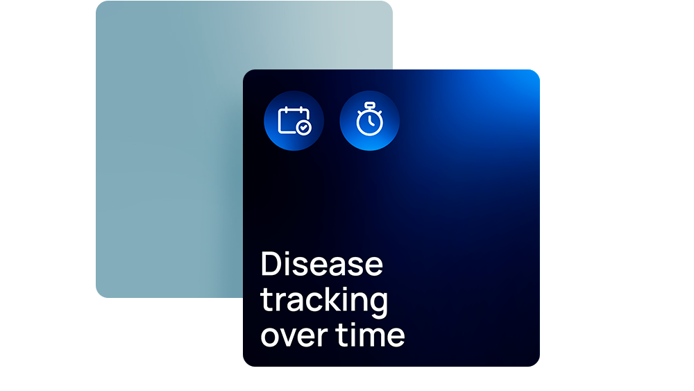
Disease tracking
Objective longitudinal reporting to track the evolution of diseases over time. -
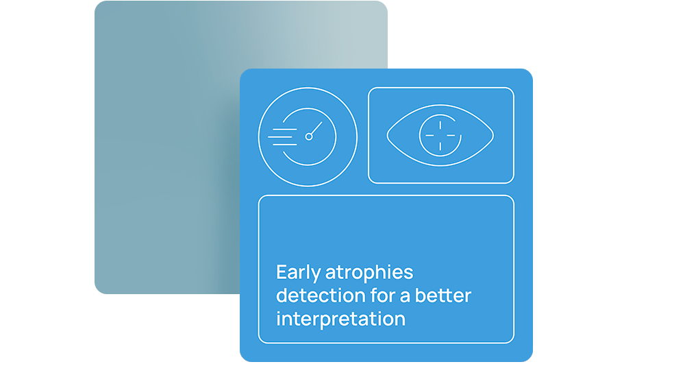
Early atrophies detection
QP-Brain® redefines brain MRI analysis by quantifying early brain atrophy and lesions for a better understanding of diseases. -
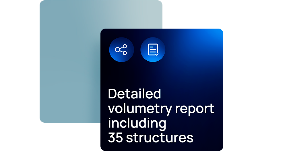
Volumetry report
QP-Brain® reports confirm visual findings with objective quantitative results. Leverages an extensive normative database. -
Regulatory status
Quibim meets international regulatory standards and is a pioneer in securing approvals across global markets. QP-Brain has received, among others, CE marking, UKCA marking, and 510(k) clearance, setting us apart in the US market.
Brain MRI device enhanced with AI technology
Quantitative analysis
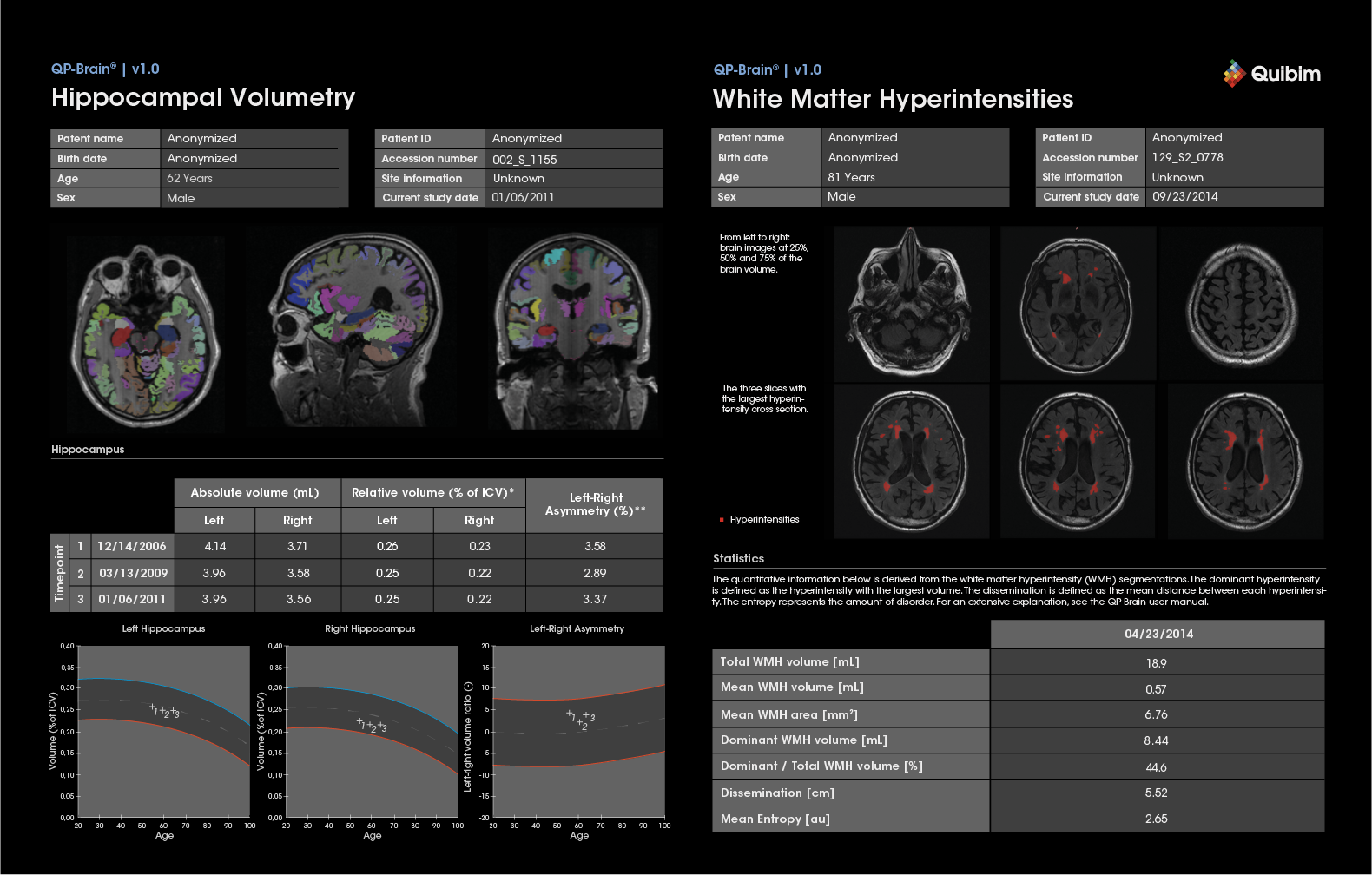
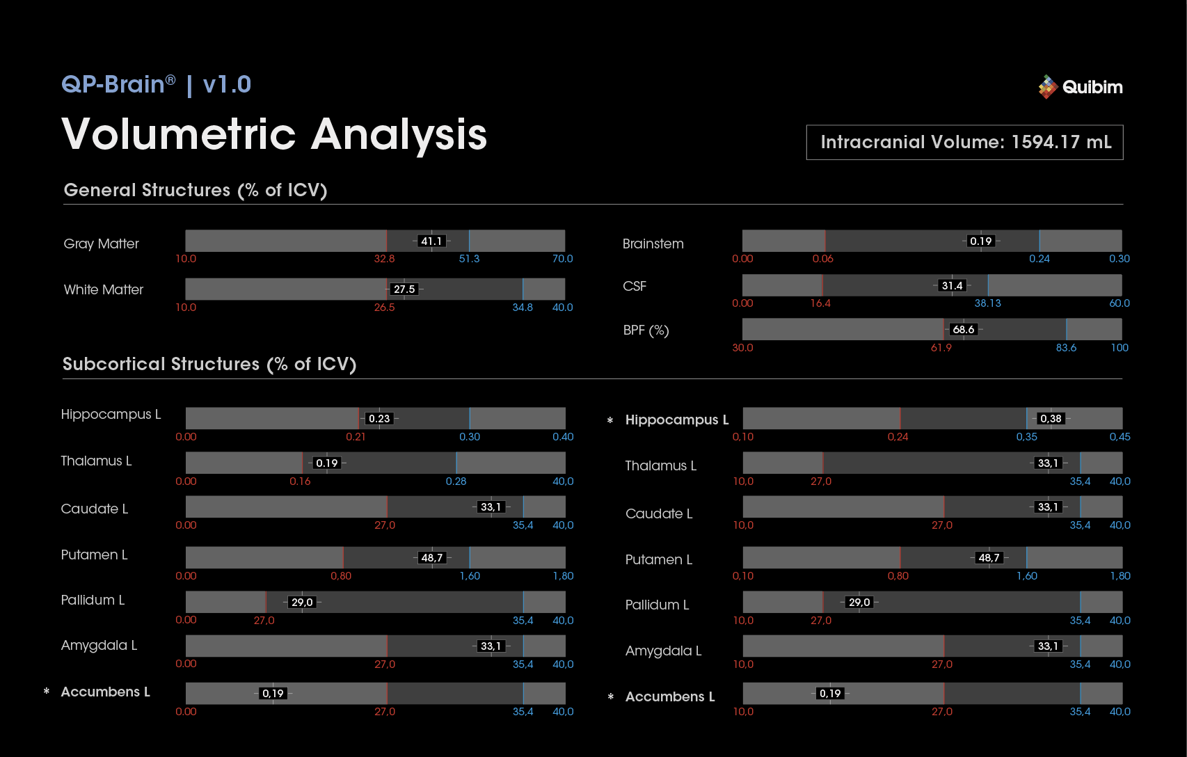
QP-Brain® offers a quantitative analysis of patients’ brain MRI, delivering automatic quantification and display of imaging findings and data, such as the volumes of key intracranial structures and WMH load/location.
Precision in brain volumetry
Outperforms in assessing central nervous system disorders by measuring absolute and relative volumes of grey and white matter, cerebrospinal fluid, and analyzing 132 distinct brain regions (L/R).
Innovative AI technology
Our patented AI ensemble excels in WMH detection and segmentation, effectively filtering out physiological hyperintensities like ependymal enhancements.
Democratizing access with standard protocols
QP-Brain® supports WMH segmentation using a 2D FLAIR input, obviating the need for 3D-T1.
Objective reporting
Confirm visual findings with objective quantitative results. Leverages an extensive normative database.
Dynamic workflow
Outputs are available for viewing in standard hospital PACS environments, appearing as an additional series in a patient folder. No additional workstations or viewing environments are necessary.
GDPR & HIPAA compliant
Installed on a hospital server behind the firewall, QP-Brain® rendering is anonymized, typically through a secure Cloud environment (AWS, Microsoft Azure).
AI brain imaging quantification software for early atrophy and lesion detection, industry-benchmarked
QP-Brain® is an AI brain image quantification software designed to improve magnetic resonance imaging (MRI) analysis of the brain, especially in the context of an aging population and increasing neurodegenerative diseases. This brain image quantification software automates de quantitative analysis of brain structure and white matter hyperintensities (WMH), providing accurate data on the volumes o different brain regions and WMH load. By comparing this data with a normative database, the software helps to detect early changes in brain atrophy and lesions, which is key for the diagnosis and monitoring of diseases such as Alzheimer’s disease.
In addition, QP-Brain® improves the efficiency of radiological reports by providing objective and quantitative data, reducing the margin of human error. With its advanced AI architecture and compliance with privacy regulations such as GDPR and HIPAA, the software facilitates access to high-quality brain analysis tools in various clinical settings. This enables better long-term monitoring of disease progression and improved diagnostic accuracy.
Automated workflow to improve patient care
AI-enhanced brain MRI device that is redefining the standards of radiological analysis
Related articles
-
Nature Biopharma Dealmakers – Quibim: empowering biopharma to turn images into actionable predictions using artificial intelligence
Read more
-

Frontiers in imaging: celebrating radiological advances with Dr. Regina Beets-Tan on International Day of Radiology
Read more
-
AI in radiology: benefits and applications
Read more