
Benefits of AI in radiology: Most significant advances and potential applications
The integration of AI technology into the field of radiology has sparked a revolution in medical imaging data. This post explores AI’s manifold benefits of AI in radiology, showcasing its wide-ranging applications and how it enhances every aspect of the radiology workflow.
AI advancements across the radiology workflow
Remarkable strides in AI technology have transformed the radiology workflow, offering significant benefits of AI in radiology across various stages: from image acquisition to final diagnosis. These AI-driven enhancements facilitate faster, higher-quality acquisitions, revolutionize the patient experience, and simplify the radiologist’s workload.
1. Enhancing image acquisition
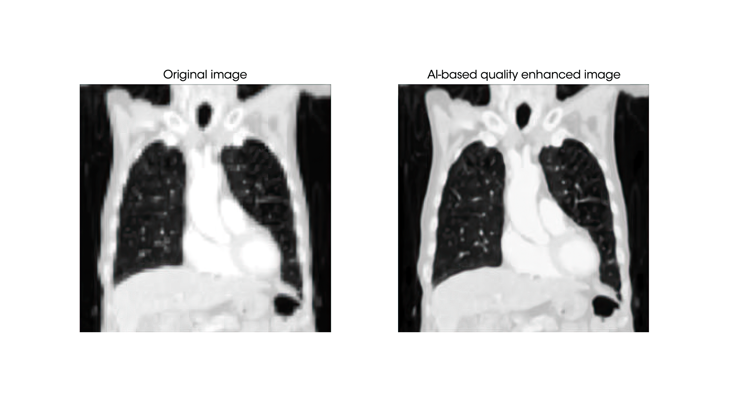
One of the primary benefits of AI in radiology is its ability to accelerate image acquisition while preserving and often enhancing image quality. We can acquire data more swiftly by utilizing AI, mitigating issues like motion artifacts caused by patient movement. This ensures a more comfortable patient experience and yields images of higher quality, optimizing diagnostic accuracy.
2. Streamlining reporting with AI
The efficiency gained in image acquisition, however, presents new challenges. Radiology departments now contend with larger volumes of images to report. Thanks to AI in radiology, this workflow can be enhanced thanks to different solutions.
- Quality enhancement algorithms: These algorithms significantly improve image quality by reducing artifacts, enhancing resolution, and augmenting the contrast between anatomical structures. This provides radiologists with clearer, more precise images for reporting.
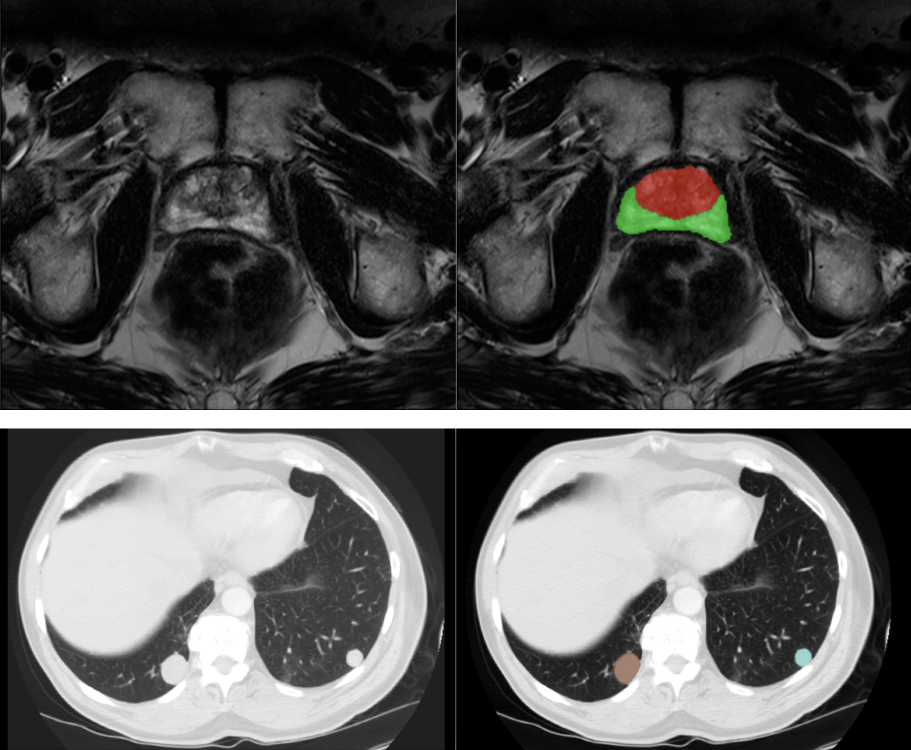
- Image segmentation algorithms: Radiologists frequently need to delineate regions of interest manually, a time-consuming endeavor. AI-powered segmentation automates this process, expediting tasks like tissue and disease characterization. Additionally, it aids in other areas, such as surgical and radiotherapy planning, where precise delineation is paramount.
- Lesion detection algorithms: Identifying anomalies within medical images can be daunting. AI assists radiologists by highlighting suspicious areas and optimizing anomaly detection and localization.
- Classification methods: When dealing with high volumes of images, another significant benefit of AI in radiology is its ability to classify and prioritize cases, improving diagnostic efficiency. They encompass two main applications:
-
- Image triage: Classification algorithms act as a virtual gatekeeper, swiftly assessing the likelihood of pathology in each image. Images deemed less likely to show abnormalities are efficiently filtered out, allowing radiologists to focus their expertise on the most relevant cases.
- Priority lists: AI-generated priority lists are a radiologist’s ally in busy clinical settings. By classifying cases based on the urgency of review, these lists ensure that critical or time-sensitive cases are addressed promptly. This not only aids in rapid decision-making but also reduces the risk of overlooking crucial findings.
Beyond Diagnosis: Prognosis with Radiomics
While AI’s role in diagnosing medical conditions is transformative, its impact extends into prognosis. In this domain, radiomics is emerging as a powerful tool for predicting clinical endpoints and patient outcomes.
Radiomics is a multidisciplinary field that extracts quantitative information from medical images. It delves deep into the textures, shapes, and other intricate patterns within images that are often invisible to the human eye. This extracted data, known as radiomic features, holds crucial insights into disease characteristics and progression.
The potential of radiomics in prognosis is broad:
- Predicting clinical endpoints: One of the core benefits of AI in radiology is its ability to analyze radiomics features and predict clinical endpoints such as disease recurrence, treatment response, and overall survival rates. AI models leverage changes in radiomics features over time to forecast a patient’s condition, aiding in more accurate predictions.
- Early intervention: Radiomics can detect subtle changes not apparent through traditional means. By identifying early signs of disease progression, radiomics empowers healthcare providers to intervene swiftly, potentially altering the course of a disease.
- Treatment monitoring: Another major benefit of AI in radiology is its ability to monitor a patient’s response to treatment through radiomics. This feedback loop allows physicians to adjust treatments in real-time, enhancing the precision and effectiveness of care.
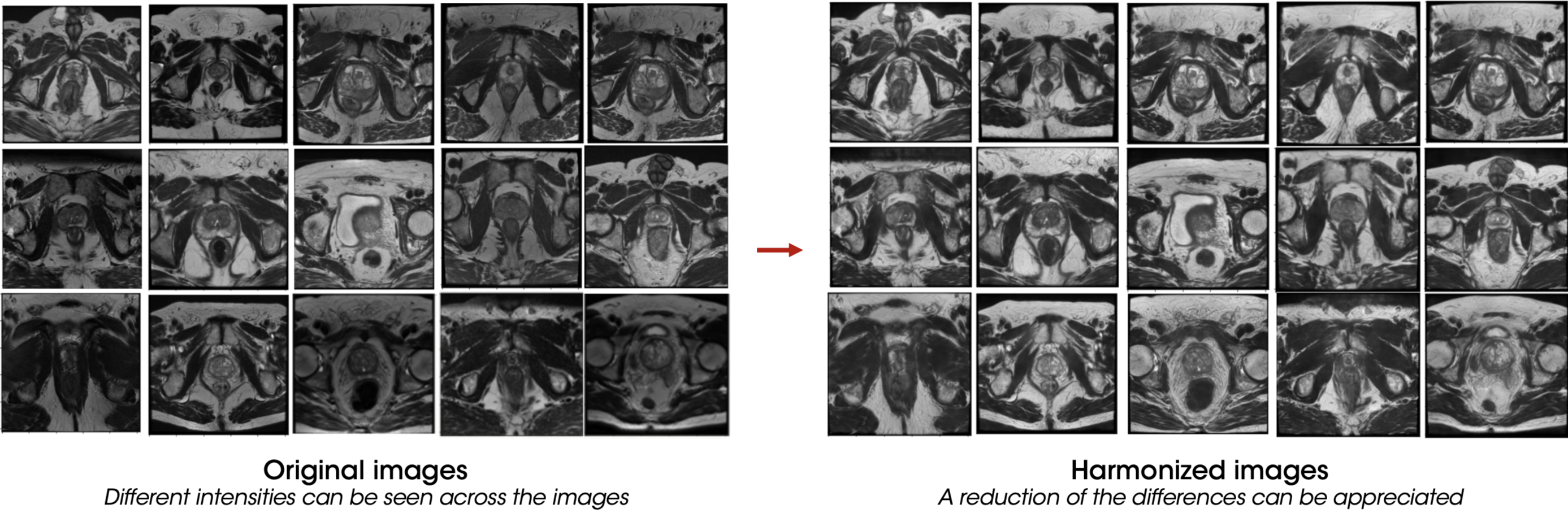
To build robust prognostic models, collecting data from various institutions is essential, considering as much variability as possible. However, the main challenge in multi-centric imaging studies lies in the heterogeneity of scanners and acquisition protocols. To address this limitation, harmonization tools are essential. AI in radiology has shown promising results in this field8, effectively reducing the variabilities commonly encountered in datasets acquired from different scanners.
Yet, the full potential of personalized medicine emerges when radiomics integrates with multiple data sources, including genomics, blood tests, and clinical data, among others. This synergy provides a comprehensive understanding of a patient’s condition, enabling the creation of personalized prognostic models. These models consider the unique biological characteristics of each patient, facilitating tailored treatment plans that are precise and effective.
Challenges and Future Directions of AI in radiology
While the benefits of AI in radiology have already revolutionized the field, several challenges and opportunities remain as the technology continues to evolve. Addressing these challenges will be key to unlocking the full potential of AI in radiology and ensuring its widespread adoption.
- Data quality and diversity: One of the core benefits of AI in radiology is its reliance on vast, diverse datasets. Ensuring data quality, diversity, and standardization remains challenging, as medical images can vary significantly regarding acquisition techniques and quality. Even though a significant improvement has been made in AI-based harmonization techniques, the future of AI in radiology necessitates collaborative efforts to create comprehensive and standardized datasets for training AI algorithms effectively.
- Ethical considerations: The ethical use of AI in radiology is paramount. Ensuring patient privacy, obtaining informed consent for data usage, and addressing issues related to bias and algorithm fairness are ongoing challenges. For AI to be widely accepted, strong ethical frameworks and regulatory guidelines will be required. These will ensure that the benefits of AI in radiology do not compromise patient rights or lead to unfair outcomes.
- Algorithm validation: Not only AI in radiology, AI algorithms need rigorous validation to ensure their reliability and safety in real-world clinical settings. Establishing standardized validation processes and frameworks will be essential to gain wider acceptance and trust among healthcare professionals and ensure the benefits of AI in radiology translate into real-world practice.
- Interoperability: Integrating AI solutions into existing healthcare systems can be complex. Radiology departments often use a variety of equipment and software systems. Future developments should prioritize interoperability to enable seamless integration and data exchange.
- Continuous learning: AI models must adapt to evolving medical knowledge and changing patient populations. Implementing mechanisms for continuous learning, where AI algorithms can update and improve over time, will be crucial to maintaining their relevance.
- Clinical adoption: While AI has shown promise, its full-scale adoption in clinical practice is still evolving. Radiologists and healthcare providers need adequate training and education to use AI tools effectively. Promoting awareness and building trust in AI-driven decision support will be critical to achieving full clinical adoption and maximizing the benefits of AI in radiology.
- Cost-effectiveness: Implementing AI in radiology is a significant consideration. Future developments should demonstrate improved patient outcomes and economic benefits, such as reduced healthcare costs and enhanced resource allocation.
Future benefits of AI in radiology holds exciting possibilities:
Looking ahead, the benefits of AI in radiology are set to expand across various areas:
- AI-augmented radiologists: AI will increasingly serve as a valuable assistant to radiologists, helping them interpret images, detect anomalies, and prioritize cases. Radiologists will focus more on complex cases and patient care, enhancing efficiency.
- Advanced prognostics: Radiomics and AI will play a more prominent role in prognosis. AI models will evolve to predict disease trajectories, optimal treatment strategies, and patient-specific outcomes with higher accuracy.
- Personalized medicine: Integrating radiomics with genomics, clinical data, and other sources will drive the emergence of truly personalized medicine. AI in radiology will facilitate the creation of highly tailored treatment plans and interventions.
- Telemedicine and remote imaging: AI-powered image analysis will enable telemedicine and remote imaging, allowing patients in underserved areas to access high-quality diagnostic services. This can revolutionize healthcare delivery worldwide.
- AI-enhanced research: AI in radiology will accelerate medical research by automating data analysis, identifying potential research subjects, and discovering new insights from vast datasets, ultimately advancing our understanding of diseases and treatments.
- Global collaboration: Collaboration among healthcare institutions, tech companies, and regulatory bodies will be critical for the responsible and effective deployment of AI in radiology. Global standards, shared datasets, and best practices will facilitate progress.
In conclusion, the future benefits of AI in radiology holds immense potential to enhance patient care further. Overcoming challenges and steering the field toward ethical, safe, and effective use of AI will require collective effort and innovation. The future promises a healthcare landscape where AI, in synergy with human expertise, enables more accurate diagnoses, personalized treatments, and improved patient outcomes.
References
- Chen Z, Pawar K, Ekanayake M, Pain C, Zhong S, Egan GF. Deep Learning for Image Enhancement and Correction in Magnetic Resonance Imaging-State-of-the-Art and Challenges. J Digit Imaging. 2023 Feb;36(1):204-230. Epub 2022 Nov 2. PMID: 36323914; PMCID: PMC9984670.
-
Jimenez-Pastor A, Alberich-Bayarri A, Lopez-Gonzalez R, Marti-Aguado D, França M, Bachmann RSM, Mazzucco J, Marti-Bonmati L. Precise whole liver automatic segmentation and quantification of PDFF and R2* on MR images. Eur Radiol. 2021 Oct;31(10):7876-7887. Epub 2021 Mar 25. PMID: 33768292.
-
Jimenez-Pastor A, Lopez-Gonzalez R, Fos-Guarinos B, Garcia-Castro F, Wittenberg M, Torregrosa-Andrés A, Marti-Bonmati L, Garcia-Fontes M, Duarte P, Gambini JP, Bittencourt LK, Kitamura FC, Venugopal VK, Mahajan V, Ros P, Soria-Olivas E, Alberich-Bayarri A. Automated prostate multi-regional segmentation in magnetic resonance using fully convolutional neural networks. Eur Radiol. 2023 Jul;33(7):5087-5096. Epub 2023 Jan 24. PMID: 36690774.
-
Lång K, Josefsson V, Larsson AM, Larsson S, Högberg C, Sartor H, Hofvind S, Andersson I, Rosso A. Artificial intelligence-supported screen reading versus standard double reading in the Mammography Screening with Artificial Intelligence trial (MASAI): a clinical safety analysis of a randomized, controlled, non-inferiority, single-blinded, screening accuracy study. Lancet Oncol. 2023 Aug;24(8):936-944. PMID: 37541274.
-
Baltruschat I, Steinmeister L, Nickisch H, Saalbach A, Grass M, Adam G, Knopp T, Ittrich H. Smart chest X-ray worklist prioritization using artificial intelligence: a clinical workflow simulation. Eur Radiol. 2021 Jun;31(6):3837-3845. Epub 2020 Nov 21. PMID: 33219850; PMCID: PMC8128725.
-
Marti-Bonmati L, Cerdá-Alberich L, Pérez-Girbés A, Díaz Beveridge R, Montalvá Orón E, Pérez Rojas J, Alberich-Bayarri A. Pancreatic cancer, radiomics and artificial intelligence. Br J Radiol. 2022 Sep 1;95(1137):20220072. Epub 2022 Jun 28. PMID: 35687700.
-
Herrero Vicent C, Tudela X, Moreno Ruiz P, Pedralva V, Jiménez Pastor A, Ahicart D, Rubio Novella S, Meneu I, Montes Albuixech Á, Santamaria MÁ, Fonfria M, Fuster-Matanzo A, Olmos Antón S, Martínez de Dueñas E. Machine Learning Models and Multiparametric Magnetic Resonance Imaging for the Prediction of Pathologic Response to Neoadjuvant Chemotherapy in Breast Cancer. Cancers (Basel). 2022 Jul 19;14(14):3508. PMID: 35884572; PMCID: PMC9317428.
-
Nan Y, Ser JD, Walsh S, Schönlieb C, Roberts M, Selby I, Howard K, Owen J, Neville J, Guiot J, Ernst B, Pastor A, Alberich-Bayarri A, Menzel MI, Walsh S, Vos W, Flerin N, Charbonnier JP, van Rikxoort E, Chatterjee A, Woodruff H, Lambin P, Cerdá-Alberich L, Martí-Bonmatí L, Herrera F, Yang G. Data harmonization for information fusion in digital healthcare: A state-of-the-art systematic review, meta-analysis and future research directions. Inf Fusion. 2022 Jun;82:99-122. PMID: 35664012; PMCID: PMC8878813.