New perspectives for cancer management: The power of combining medical imaging with real–world evidence
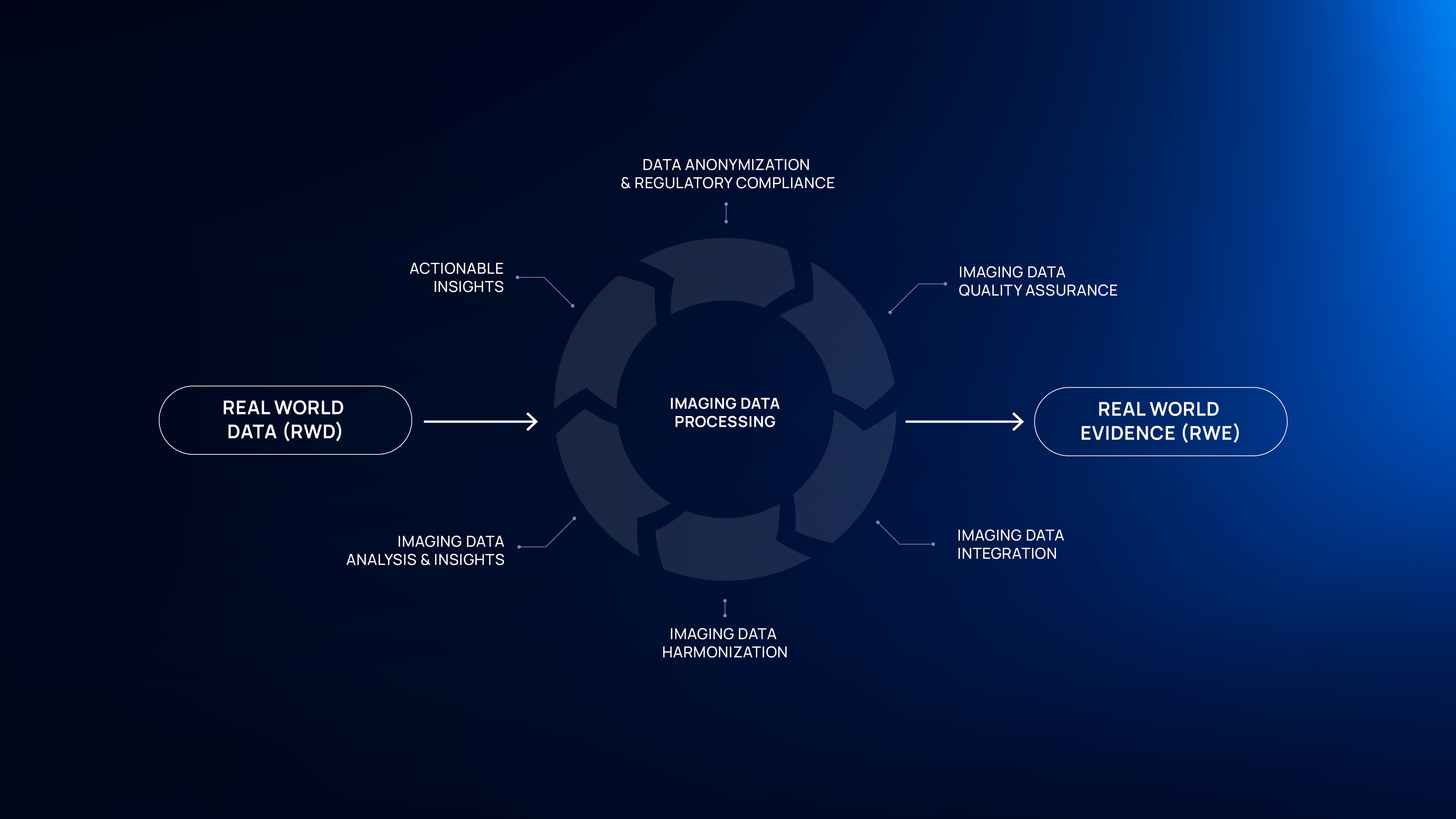
The landscape of cancer management is undergoing significant transformation with the integration of medical imaging and real-world evidence (RWE). Maximizing real-world data (RWD) to produce real-world evidence (RWE) has become an increasingly important approach, supplementing traditional clinical trials. This approach is gaining significant traction, particularly in the field of oncology, share its applications are expanding rapidly, reflected in a rising number of studies and its growing role in regulatory decisions over the past few years1.
In parallel, biomedical imaging is playing an ever more important role in all phases of cancer management, from prediction and screening to therapy response and palliation2. Together, these two evolving fields are reshaping cancer research by offering complementary insights, where RWD provides real-world context, and imaging contributes crucial visual data for patient-specific diagnosis and treatment planning.
Understanding real–world evidence in cancer research management
In the context of cancer management, RWD refers to health information collected outside the framework of traditional randomized controlled trials, often derived from routine clinical practice. When properly analyzed, this data can be transformed into RWE, offering valuable insights into the effectiveness and risks of therapeutic interventions as experienced in everyday clinical environments, as opposed to controlled trial settings3 RWD can come from numerous sources, with patient registries, electronic health records (EHRs), and administrative claims being among the most prevalent3.
RWE offers several key advantages for cancer management. Firstly, RWD encompasses larger and more diverse datasets, enhancing the generalizability of research findings to a broader population. Secondly, since RWD is often collected before the research begins, studies can be completed in a matter of weeks or months rather than years. Additionally, RWD studies typically involve fewer procedures and lower costs and sometimes, they represent the only feasible option for investigating treatments for rare diseases3. Despite these unquestionable benefits, still several challenges need to be overcome, including the homogenization in regulation around data access and data privacy.
The role of medical imaging in cancer management
Medical imaging is a cornerstone of cancer management, playing a crucial role in early detection, diagnosis, staging, and monitoring treatment response.
In cancer management, imaging serves several essential functions:
Advanced imaging techniques now enable early cancer detection, offering more effective treatment options and better recovery prospects. Additionally, screening programs have enhanced early-stage detection. Imaging technologies such as magnetic resonance imaging (MRI), CT, and positron emission tomography (PET) scans provide precise tumor details, helping to differentiate between benign and malignant tissue. For example, mammography has reduced breast cancer mortality by 60%4, and low-dose CT scans have cut lung cancer deaths by 20%5. Moreover, imaging is crucial for staging cancer, determining its spread, guiding treatment decisions, ensuring tailored and effective therapeutic approaches.
Synergizing medical imaging and real-world evidence for cancer management
The integration of medical imaging with RWE brings significant advantages to cancer management, enhancing personalization, detection, and long-term monitoring of cancer treatments. These are some of the advantages that this approach may provide:
- Enhanced treatment personalization: By combining RWE with imaging data, researchers can analyze large and diverse populations, helping to better understand how various treatments perform across different demographics. This integration enables a more personalized approach to cancer treatment, tailoring interventions to individual patient profiles based on real-world outcomes.
- Improved early detection and monitoring: When paired with RWE, medical imaging can help track how patients respond to treatment over time, offering insights into real-world scenarios where clinical trial data might be limited. This allows for earlier intervention and adjustments to treatment, improving patient outcomes.
- Better understanding of long-term outcomes: Clinical trials typically have controlled conditions and limited follow-up durations. By integrating medical imaging into RWE, researchers can assess long-term outcomes of cancer treatments, monitoring disease progression or remission over extended periods. This helps to understand how treatments work in real-life conditions, including in populations not well-represented in trials, such as the elderly or those with comorbidities.
- More accurate staging and prognosis: Imaging provides detailed insights into the size, location, and spread of tumors, which are essential for staging cancer and guiding treatment decisions. Coupling this with RWE allows researchers to track how different stages of cancer progress in real-world settings, leading to better prognosis predictions and more effective treatment planning.
- Validation of clinical trial results: RWE and medical imaging can be used to validate clinical trial results in broader populations. Imaging data from real-world patients can confirm whether treatment effects observed in trials are consistent when applied in everyday clinical practice, helping to solidify the evidence base for treatment protocols.
- Accelerating drug development: RWE, supported by imaging data, can be used to support regulatory approvals by providing evidence of a drug’s effectiveness in routine clinical use. This can shorten the timeline for drug development and bring new treatments to the market more quickly, benefiting patients who need innovative therapies.
- Optimized resource allocation: By analyzing real-world imaging data in conjunction with patient outcomes, healthcare providers can better allocate resources, such as determining which imaging techniques are most cost-effective or identifying patient populations that would benefit the most from specific imaging technologies.
As the integration of real-world evidence and medical imaging continues to evolve, it will play an increasingly critical role in cancer management. By harnessing the power of real-world data alongside imaging insights, healthcare providers, researchers, and regulators are better equipped to optimize patient outcomes, enhance treatment protocols, and expedite the development of innovative therapies.
The synergy between medical imaging and real-world evidence offers new, powerful tools for advancing cancer management. By embracing this innovative approach, the medical community can better respond to the challenges of cancer care, delivering more personalized, effective, and timely treatments to patients worldwide.
References
- Castelo-Branco, L. et al. (2023) ESMO Guidance for Reporting Oncology Real-World evidence (GROW). Annals of Oncology, Volume 34, Issue 12, 1097 – 1112. DOI: 10.1016/j.annonc.2023.10.001
- Fass, L. (2008). Imaging and cancer: A review. Molecular Oncology, 2(2), 115-152. DOI: 10.1016/j.molonc.2008.04.001
- Saesen, Robbe, et al. (2023) Defining the role of real-world data in cancer clinical research: The position of the European Organisation for Research and Treatment of Cancer. European Journal of Cancer, Volume 186, 52 – 61. DOI: 10.1016/j.ejca.2023.03.013
- Duffy SW, et al. (2020). Mammography screening reduces rates of advanced and fatal breast cancers: Results in 549,091 women. Cancer. Jul 1;126(13):2971-2979. DOI: 10.1002/cncr.32859
- The National Lung Screening Trial Research Team. (2011) Reduced Lung-Cancer Mortality with Low-Dose Computed Tomographic Screening. New England Journal of Medicine. 365(5): p. 395-409. DOI: 10.1056/NEJMoa1102873