Improving MRI workflows with AI-enhanced advanced medical imaging technology
Medical imaging has become so important in the modern care environment that one can only think of providing clinical care with visual information on human internal structures. This technology supports diagnosing, monitoring, and treating various medical conditions. Medical imaging helps diagnose, monitor, and cure different conditions. Imaging modalities such as X-ray, CT, MRI, PET, and ultrasound are important in diagnosing diseases related to cancer, cardiac disorders, and neurological disorders. These imaging methods improve our understanding of normal and abnormal anatomy, assisting clinicians in making informed decisions.
Recently, medical imaging technology integrated with AI reimagines workflows, enhancing diagnoses’ speed, accuracy, and efficiency. These recent improvements have hastened diagnoses, improved their accuracy and effectiveness at higher resolutions in real-time, and even allowed AI-assisted personalized treatment planning. Also, AI will help overcome some of the other challenges, such as those related to radiation exposure and complex image interpretation. With the continuous rise in these technologies, there is the capacity to bring greater efficiency into health processes by reducing errors and having more correct care, thus leading to better management of complicated diseases and disorders.
Advances in medical imaging technology
Today’s advanced imaging technologies offer precise, non-invasive diagnostic solutions for a wide range of medical needs. Computed Tomography (CT) uses X-rays from multiple angles to produce detailed cross-sectional images, effectively detecting cancers, monitoring tumor progression, and diagnosing cardiovascular issues. Innovations like volumetric quantitative CT (QCT) and high-resolution CT have greatly improved bone imaging and osteoporosis management applications.
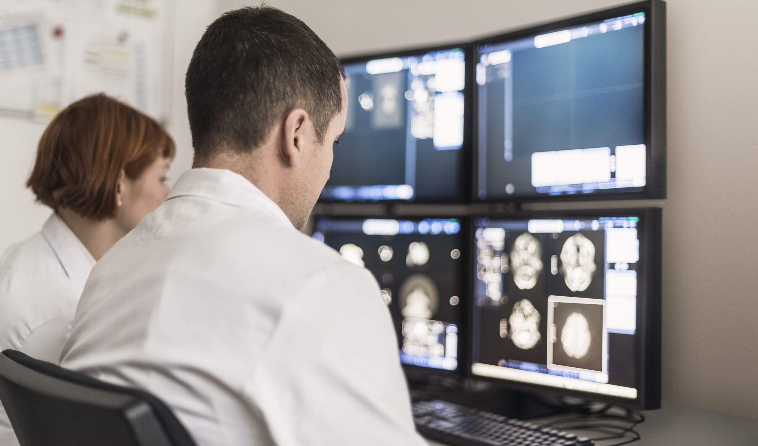
Magnetic Resonance Imaging (MRI) applies a magnetic field and radiation to obtain the maximum possible structural resolution of soft tissue anatomy. It is an excellent imaging tool for neurological conditions such as multiple sclerosis, as well as diseases of the heart. Functional MRI (fMRI) and Magnetic Resonance Angiography (MRA) broaden MRI’s capabilities, allowing for detailed studies of brain functions and vascular systems.
Nuclear imaging techniques, including Positron Emission Tomography (PET) and SPECT, provide unique insights into metabolic activity and blood flow, which is invaluable for diagnosing and treating oncology, cardiology, and neurology. In hybrid PET/MRI, each technique brings strengths that enhance precision for tumor analysis and other diseases with complex medical conditions.
Digital mammography has improved the early detection of breast cancer, offering higher-resolution images compared to traditional film mammography. This is particularly helpful in young women with dense breast tissue, as it increases diagnostic accuracy and allows for timely interventions.
Ultrasound sonography remains a versatile tool. It uses high-frequency sound waves to image soft tissues and organs. Its applications span obstetrics, cardiology, and abdominal imaging, making it a fundamental part of routine and emergency medical diagnostics.
The role of AI in medical imaging technology
Artificial intelligence is transforming medical imaging technology by improving image analysis accuracy, speed, and efficiency, making it a vital part of contemporary healthcare. It is used in detecting abnormalities and automating reports, among others, with machine learning and deep learning technologies.
AI-driven systems, such as convolutional neural networks (CNNs), can recognize complex patterns to diagnose conditions like lung cancer, brain tumors, and cardiovascular diseases at an early stage. AI enhances visualization by evaluating image quality, extracting imaging biomarkers for personalized treatment planning, and enabling precise lesion segmentation to improve surgical and radiotherapy planning.
Additionally, it integrates imaging studies with electronic health records to provide integrated knowledge for multidisciplinary decisions. In oncology, AI is especially good at detecting lung and breast cancers. It accurately detects pulmonary nodules and tumors in dense breast tissue while tracking tumor size and texture changes to assess treatment effectiveness.
Despite these advancements, using AI in medical imaging encounters challenges such as the necessity for large, diverse datasets, regulatory hurdles, and ethical considerations. Nevertheless, ongoing research is working to tackle these issues and create robust and equitable AI-driven solutions.
References
TechTarget. (n.d.). Medical imaging. TechTarget. https://www.techtarget.com/whatis/definition/medical-imaging
U.S. Food and Drug Administration (FDA). (2023, April 17). Medical imaging. U.S. Food and Drug Administration. https://www.fda.gov/radiation-emitting-products/radiation-emitting-products-and-procedures/medical-imaging
Saha, G. B., & Prabhakar, A. (2021). Advanced medical imaging techniques. PubMed Central (PMC). https://pmc.ncbi.nlm.nih.gov/articles/PMC9192206/#:~:text=in%20imaging%20techniques.-,Computed%20tomography%20(CT)%2C%20positron%20emission%20tomography%20(PET),in%20advanced%20medical%20imaging%20techniques.