Seeing beyond a prostate cancer case – A human story
Blog
Tags:
Cancerprostate cancer
A 76-year-old male patient visited the Urology Department after undergoing a blood test and presenting an elevated prostate-specific antigen (PSA) of 7ng/mL, not too high to confirm malignancy, not too low to wait until the next follow-up. Due to the borderline PSA value, a magnetic resonance (MR) examination was indicated to rule out adenocarcinoma or other possible lesions in the prostate gland. A multiparametric prostate MRI protocol was followed acquiring a weighted T2 (T2w), a diffusion-weighted imaging (DWI) and a dynamic contrast-enhanced (DCE), crucial for detecting abnormalities in prostate segments MRI.
The radiological report showed a dominant index lesion with a focal T2 signal abnormality located in the right peripheral zone (PZ). The DWI sequence demonstrated restricted diffusion, consistent with a PI-RADS 4 lesion. Regarding the standard blind biopsy, a prostate adenocarcinoma was detected in a total of six cylinders. Therefore, the patient participating in the study had a Gleason Score of 7 (4+3), which means a high risk of clinically significant prostate cancer.
All the above findings were detected using conventional methods and the qualitative radiological reading. Accurate segmentation prostate analysis requires highly specialized knowledge of prostate physiology and pathology. Even when skilled radiologists are present, the sheer volume of MRI data makes interpretation resource-intensive, leading to delays in prostate cancer case study assessments. In this case, the patient had to wait one month before receiving a confirmed diagnosis and starting treatment.
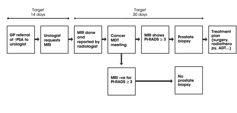
Current prostate cancer pathway
Is there a chance to streamline prostate cancer diagnosis and improve segmentation prostatique?
Quibim’s QP-Prostate® is an FDA 510(k), UKCA, and CE mark-cleared AI solution integrated into the hospital environment to accelerate prostate MRI reading and provide automated prostate segmentation. The solution offers:
With the integration of QP-Prostate® solution in that hospital, the clinical flow of this man of 76 years of age would have been reduced by 10-20 days, subsequently, his treatment plan would have started sooner.
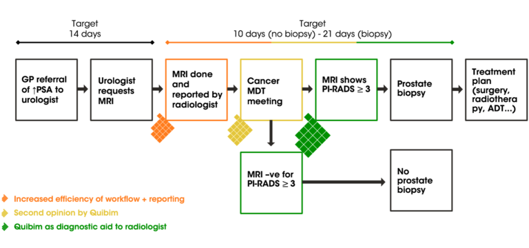
Quibim in current prostate cancer pathway
QP-Prostate® offers a fundamental change in the segmentation prostatique and prostate MRI diagnostics, increasing accuracy and efficiency and speeding up the patient journey.
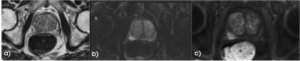
Figure 1. Representative images from the MR sequences where a) the T2w axial image, b) the DWI sequence and c) the DCE sequence.
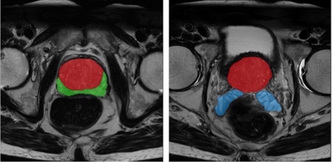
Figure 2. T2w MRI with the anatomical segmentation of the prostate gland overlayed (two different anatomical slices). The color map used is red for TZ + CZ, green for PZ and blue for seminal vesicles.
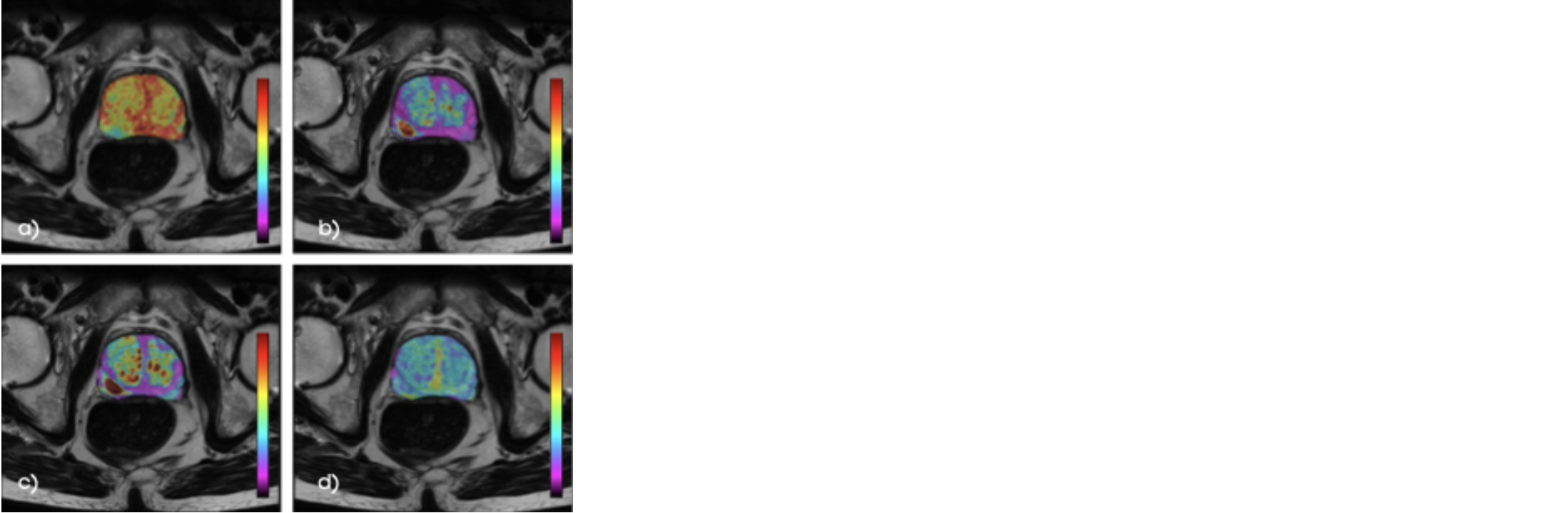
Figure 3. ADC (apparent diffusion coefficient) (a), Ktrans (volume transfer constant from the plasma compartment to the extravascular extracellular space) (b), kep (rate constant for transfer between extravascular space and the blood compartment) (c) and ve (volume of extracellular space per unit volume of tissue) (d) maps automatically calculated and shown as an overlay to the T2w, where red is associated with a high value and blue and purple with a low value.
The implementation of automated prostate segmentation and AI-driven prostate MRI analysis significantly enhances segmentation prostate accuracy, optimizes prostate cancer case study workflows, and reduces delays in diagnosis. QP-Prostate® transforms the way prostate segments MRI are analyzed, ultimately leading to better patient care and timely treatment decisions.