QP-Prostate®
Discover QP-Prostate®
Prostate cancer is the second most common cancer in men, representing a significant public health concern. While MRI scans are essential for early detection, the growing demand has surpassed the availability of radiology experts, with diagnostic delays and inconsistencies in interpretation as direct consequences. Only a minority of the medical community consistently follows PI-RADS v2.1 guidelines.
QP-Prostate introduces enhanced diagnostic capabilities, streamlining radiologists’ workflows by automatically ensuring compliance with PI-RADS v2.1 guidelines, accurately segmenting the prostate gland, and efficiently identifying suspicious lesions. These advancements enable radiologists to provide faster and more accurate assessments, directly improving patient care.
Aggressive cancer detection and diagnosis
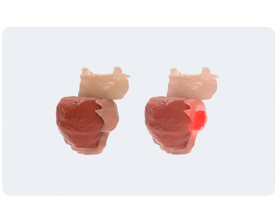
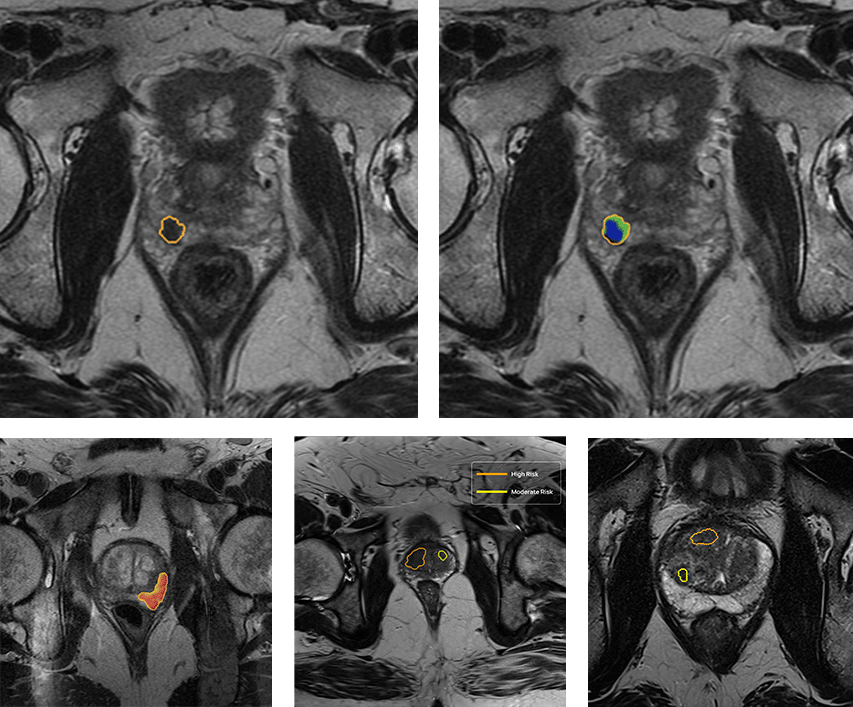
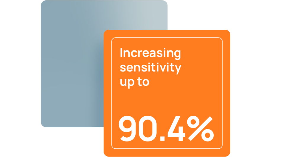
QP-Prostate is setting a new standard for accuracy, speed, and diagnostic precision. Its AI algorithm identifies and stratifies by likelihood intermediate and high-grade aggressive prostate cancer lesions, and provides a segmentation of the prostate. This allows Health Care Professionals to improve their diagnostic accuracy and fusion biopsy planning.
AI-QUAL™: Automated Prostate Image Quality Assessment
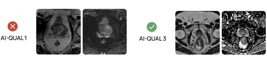
Quibim’s AI-QUAL™ automates the assessment of prostate MRI image quality based on the PI-QUALv2 guidelines. This tool streamlines workflows and supports diagnostic confidence.
Key benefits of AI-QUAL include:
- Automated Quality Scoring: Eliminates manual checks, saving time and reducing subjectivity.
- Standardized Reporting: Delivers consistent quality score based on PI-QUAL v2 guidelines.
- Artifact Detection: Identifies key image-degrading factors such as rectal gas distortion and metal artifacts.
- Diagnostic Confidence: Ensures radiologists work with the highest quality images.
- Seamless Integration: Designed to fit effortlessly into existing radiology workflows.
Your AI solution for advanced prostate cancer detection

QP-Prostate: a set of enhanced diagnostic capabilities
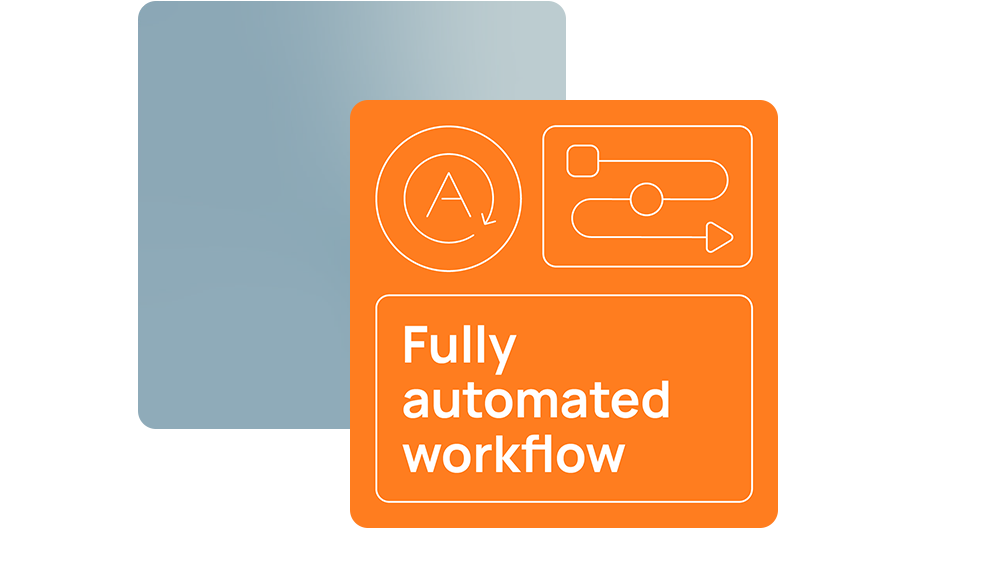
Fully automated workflow
All analysis outputs are directly sent back to the hospital PACS for their visualization without disrupting radiology workflows.
AI-Based automatic lesion detection
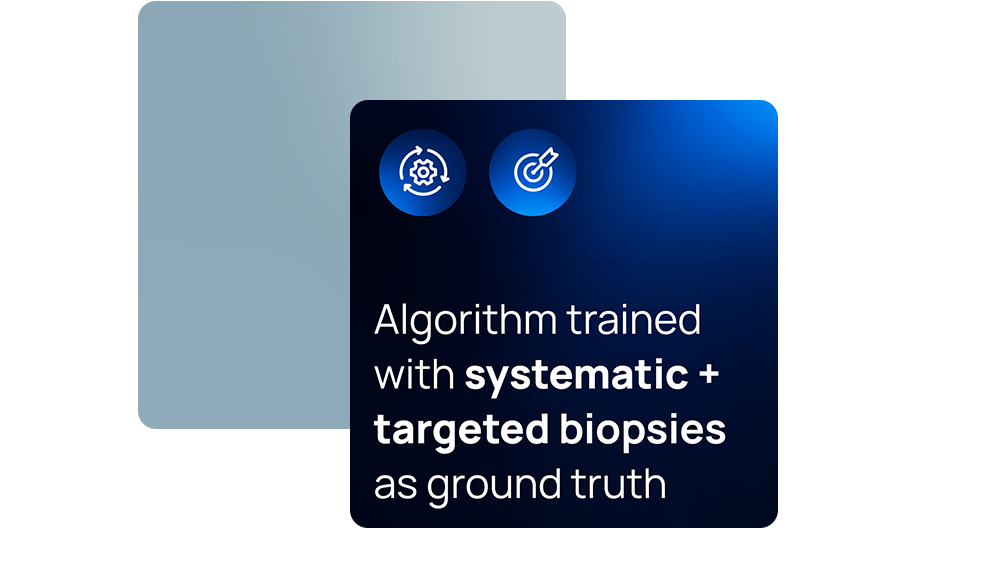
Our AI algorithm, trained with pathology outcomes as ground truth, is designed to efficiently detect clinically significant prostate cancer lesions using biparametric inputs (T2w, DWI and ADC), outperforming competitors in detection rate and speed. These AI algorithms are intended to elevate radiologists’ diagnostic accuracy with automated detection of biopsy-proven, clinically significant prostate cancer lesions.
AI-QUAL™: Automated Prostate Image Quality Assessment
AI-QUAL™ automates the assessment of prostate MRI image quality based on the PI-QUALv2 guidelines. This tool streamlines workflows and supports diagnostic confidence.
Precision in segmentation
QP-Prostate’s AI algorithm automatically identifies the prostate anatomy and segments the full prostate gland with market-leading performance (88% DSC1), as well as the seminal vesicles, to assist in PSA density calculations. The results can be directly exported for fusion biopsy procedures.
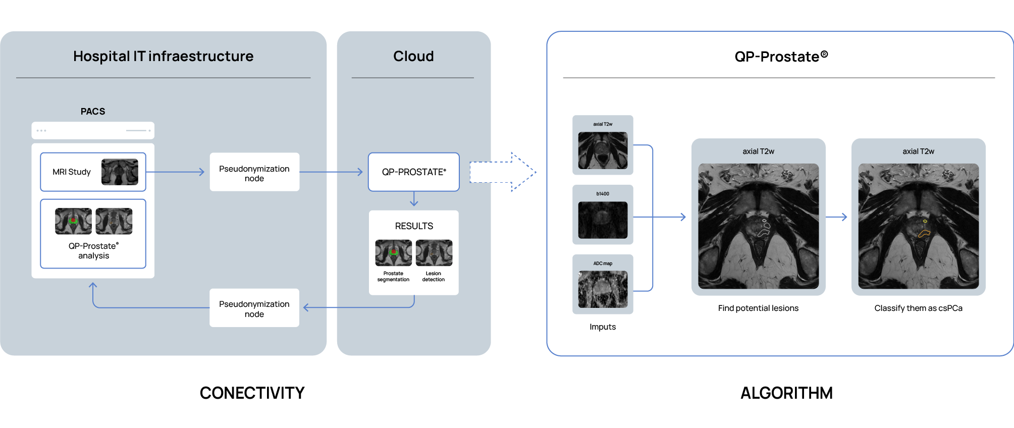
How does it work?
Clinical cases using QP-Prostate


Changing the narrative in prostate diagnosis thanks to maximum accuracy in prostate MRI interpretation
In short, QP-Prostate leverages advanced artificial intelligence image processing techniques to offer a state-of-the-art approach to prostate MRI interpretation. This image processing software is compatible with standard workstations and allows for the visualization and analysis of DICOM data by qualified professionals.
Developed by Quibim, QP-Prostate excels in automating the segmentation of critical prostate regions, including the transition zone, peripheral zone and seminal vesicles. By streamlining these complex processes, the software not only aids in the interpretation of prostate MRI images, but also enables more accurate diagnoses. This combination of sophisticated AI-based analysis and user-friendly implementation makes QP-Prostate an invaluable tool in the hands of healthcare professionals, ensuring better patient outcomes through more reliable and detailed assessments.
References
-
Jimenez-Pastor A, et al. Eur Radiol. 2023;33(7):5087-5096.
-
Sánchez Iglesias Á, et al. Cancers (Basel). 2023;15(16):4163.
*Pilot study conducted in 2023 at Massachussets General Hospital.