AI-Powered Imaging to Accelerate Oncology Drug Development
Optimized patient selection and cohort enrichment via multi-omic biomarker profiling
With capabilities across image standardization, AI-powered lesion detection and segmentation, and AI-based predictive imaging biomarkers, Quibim’s platform enables optimized trial design, expedites decision-making, and enhances the development of personalized treatments.
By combining quantitative imaging analysis extracted from SoC images with multi-omic biomarker profiling, we not only optimize patient selection and cohort enrichment but also, reveal tumor heterogeneity, microenvironment traits, and treatment response patterns. Used alone or with patient data, these biomarkers help identify those most likely to benefit from targeted or immuno-oncology therapies—enhancing trial efficiency through biologically driven cohort enrichment and accelerating time to outcome.
Image harmonization preprocessing for multi-site reproducibility
Ensure high-quality, standardized insights from medical images across sites and scanners with advanced harmonization
We often encounter variability in real-world imaging data due to differences in acquisition protocols and scanner types, leading to significant shifts in image intensity. These inconsistencies hinder traditional interpretation, reduce AI model robustness, and compromise the reproducibility of imaging biomarkers—particularly in MRI.
Harmonization is a critical pre-processing step to enhance AI model generalizability and ensure reproducibility in large-scale studies.
Quibim’s harmonization pipeline standardizes images across scanners and protocols, ensuring consistency and improving AI reliability in multi-site clinical trials.
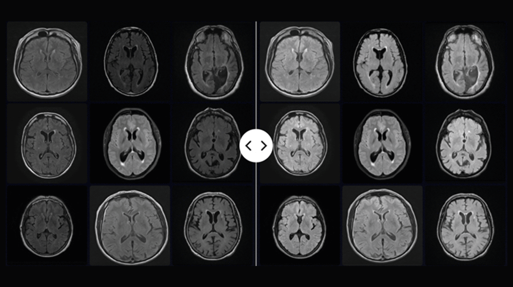
Dataset of MRI images pre- (left) and post-harmonization (right)
Automated organ and lesion detection and segmentation
Accelerate imaging workflows and reduce variability in high-throughput oncology studies.
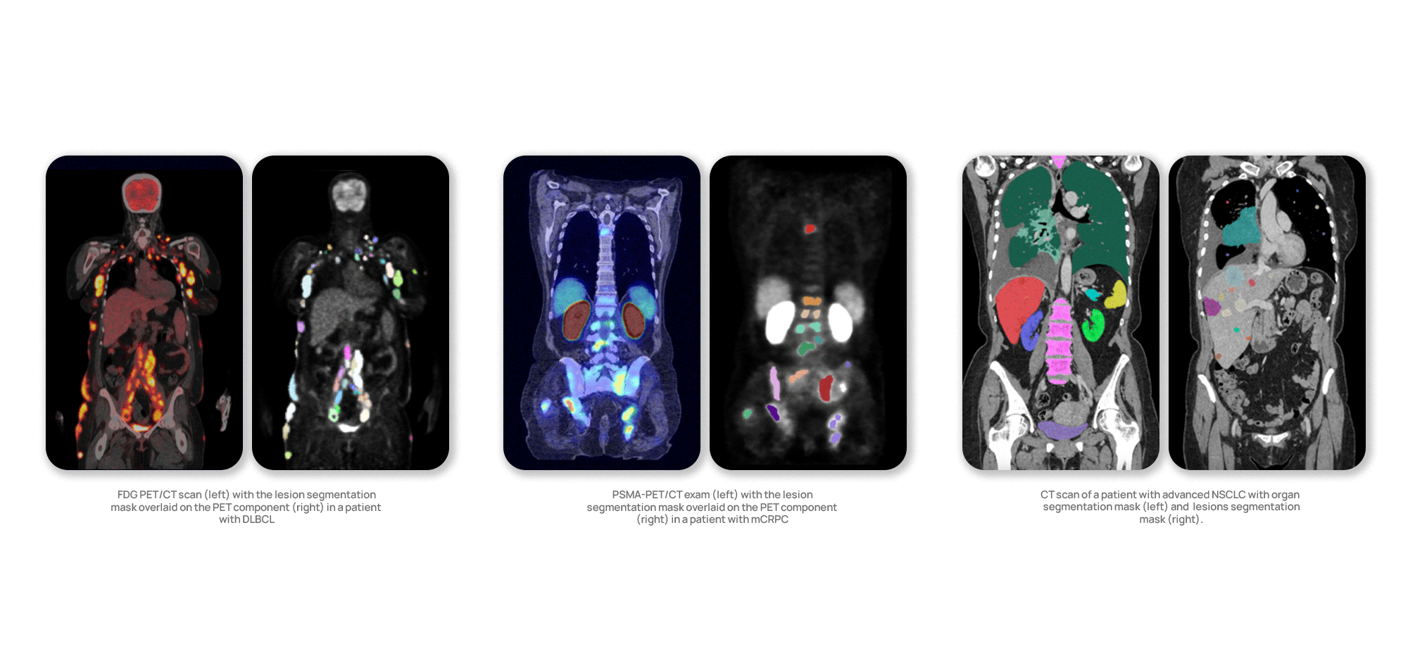
Our validated segmentation algorithms identify, segment and measure organs and lesions across PSMA-PET/CT, FDG PET/CT, CT and MRI scans. These tools drastically reduce the manual effort traditionally made by readers and provide consistent, reproducible results across diverse datasets.
Validated, fully automated segmentation across multiple organs is overcoming this barrier. Ongoing development aims to meet and exceed clinical trial standards for performance, scalability, and precision.
Quantitative lesion characterization and tracking
1. AI-supported lesion-assessment tool
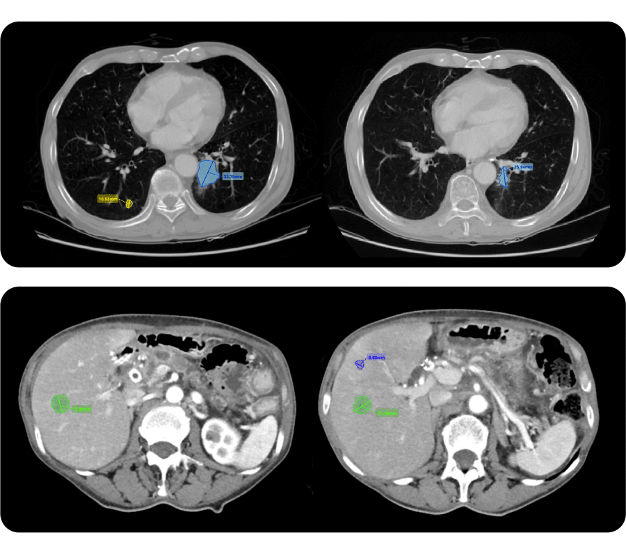
Automatically detected, segmented, and measured target lesions of a patient with advanced NSCLC at baseline [left] and 1st on-treatment scans [right]
Quibim’s tools automatically identify, segment, measure, and track lesions throughout the clinical trial. By streamlining these processes, faster and less variable radiological assessments are enhanced to achieve more consistent interpretations. Experts can also manually segment or adjust automated results within an integrated DICOM viewer, supporting RECIST 1.1 and its variations without compromising workflow efficiency.
Features include:
-
Tumor burden quantification across multiple organs, with ongoing expansion
-
Tumor growth kinetics for retrospective analysis and predictive simulation
-
Tumor heterogeneity assessment via radiomics
Benefit: Robust monitoring of treatment response and tumor progression.
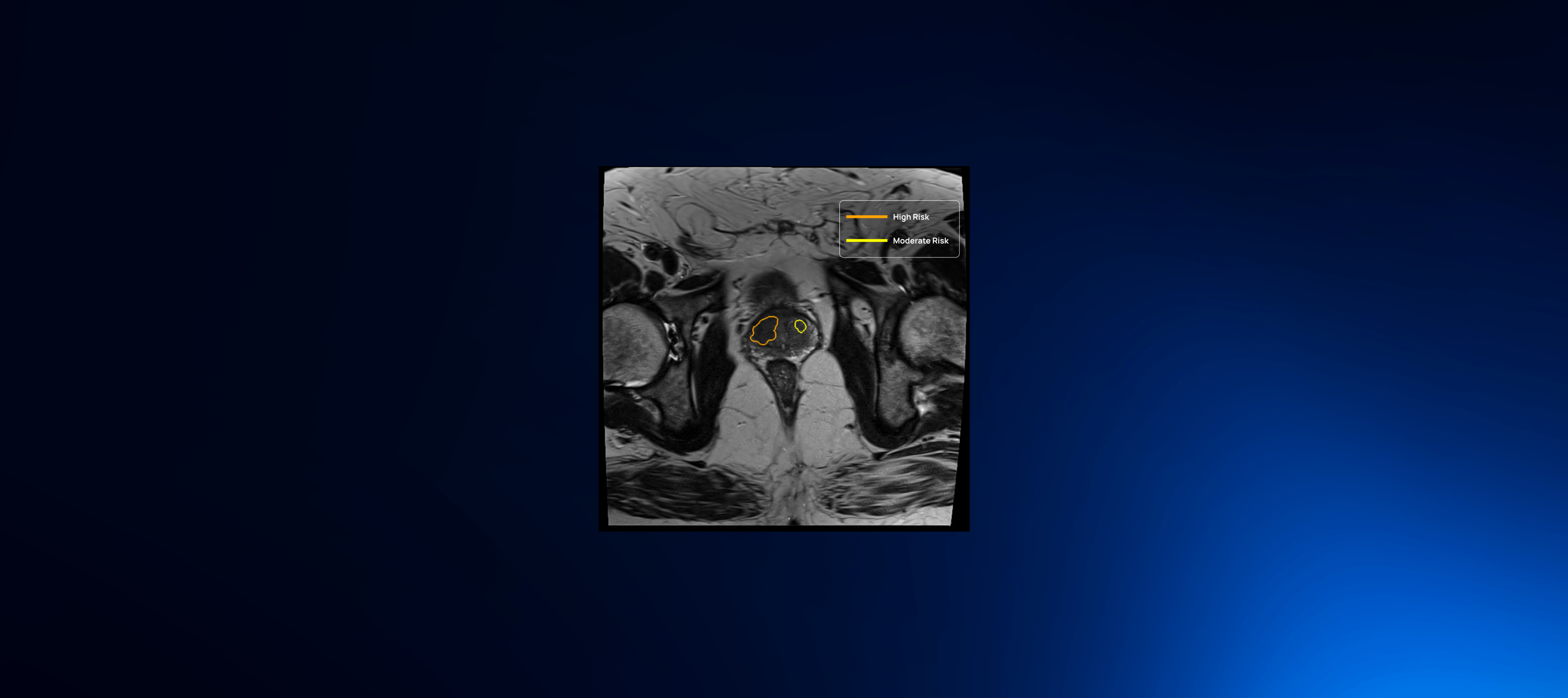
2. Whole-body tumor burden assessment
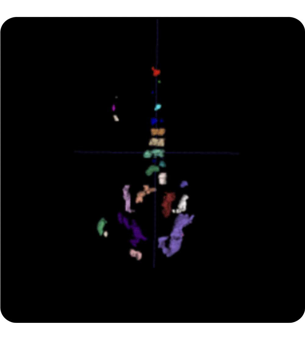
3D segmentation mask from a [18F] PSMA PET/CT scan in a patient with mCRPC. Metrics include tumor count, organ-specific and total tumor volume (TTV). For molecular imaging (e.g., PSMA PET, FDG PET), uptake-related metrics are also assessed.
3. Tumor growth kinetics
Retrospective assessment and predictive simulations for individual lesions and tumor burden evolution provides insights into tumor evolution under multiple treatment arms. This capability allows researchers to model and compare treatment outcomes with unprecedented precision.
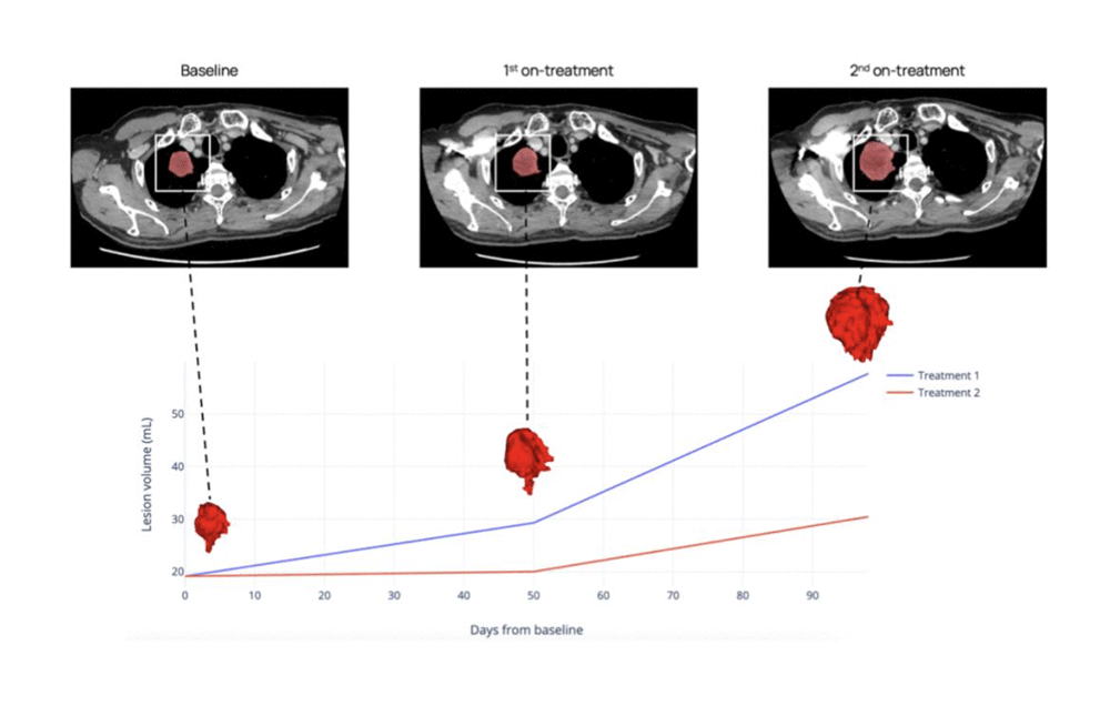
CT scans of a patient with advanced NSCLC showing target lesion evolution at three time points. This conceptual simulation illustrates how tumor growth kinetics and delta-radiomics can be assessed across treatment regimens. Not based on an actual predictive model.
4. Tumor heterogeneity assessment
Entropy and other advanced radiomic features extracted from medical images help quantify intra-lesion variability; crucial for understanding tumor aggressiveness and predicting treatment response.

Intra-lesion heterogeneity quantified using entropy from a DWI MRI scan. Texture features reveal microscopic heterogeneity beyond visual assessment. High entropy often indicates more aggressive tumors, while low entropy suggests better treatment response. Temporal changes may serve as early biomarkers of therapeutic effect.
5. Feature-based models
Feature-based models for the extraction of thousands of tissue-related variables (including radiomic features, deep features & imaging biomarkers).
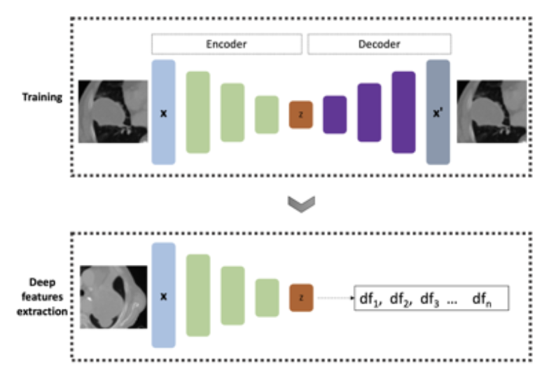
Advanced deep feature extraction from a tumor bounding box and its microenvironment using an encoder-decoder architecture
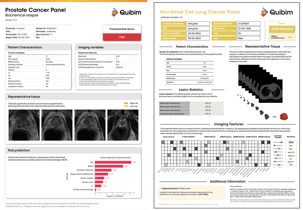
Scalable imaging biomarker panels for precision medicine
We extract high-dimensional features—including radiomic, deep features, and imaging biomarkers—from organ- and lesion-level data to build tailored panels for diagnostic, prognostic, and predictive use. These panels support cohort enrichment and patient stratification. Quibim’s biomarker panels enable detailed disease characterization, response tracking, and treatment assessment. We are expanding into mutational imaging classifiers—AI models that non-invasively predict molecular features, such as EGFR mutation status in NSCLC. By combining advanced image analysis with predictive modeling, Quibim is shaping the future of precision medicine.

Find all these functionalities in QP-Insights, an end-to-end imaging platform
The new generation advanced platform for the management, storage and analysis of large-scale multi-omics data and medical images in clinical studies and research projects.