The power of accurate segmentations in AI-driven solutions for medical imaging
Enhancing medical AI segmentation for precision and innovation
The medical AI segmentation process involves precisely identifying and delineating a region of interest (ROI) within an image. In medical image segmentation AI, these ROIs may include lesions, organs, healthy tissues, or structures, delineated in one or multiple imaging modalities, such Magnetic Resonance Imaging (MRI), Computerized Tomography (CT) or Positron Emission Tomography (PET), among others.
Precision at Quibim: Advancing AI-Driven solutions
At Quibim, the precision in medical AI segmentation is paramount. Our accurate solutions for medical training and research provide high-quality segmentation results that serve as key elements in characterizing the aggressiveness and heterogeneity of tumors, ultimately impacting patient evolution. These segmentations are also crucial for developing AI-driven solutions capable of making predictions at both lesion and patient levels.
For this reason, a standardized segmentation process is systematically implemented in every study we manage at our company, leading to the definition of reproducible and standard protocols refined to each specific use case. Not only the segmentation of a lesion is important, but also its surrounding organs and tissues. For instance, in AI-driven solutions for overall survival (OS) prediction in lung cancer, not only is the lesion ROI considered, but also nearby structures such as lungs, heart, bone/calcium, liver, metastasis, among others.
Challenges in medical image segmentation AI
There are many factors that significantly affect the accuracy of medical AI segmentation, like spatial and contrast resolution, image noise as well as variability in the shape, texture and perception of pathologies1. This highlights the importance of conducting quality controls before embarking on any image processing.
Segmentation training for AI models requires precise labels or annotations to ensure reliable outcomes. It consists of a set of tasks to select images that have enough diagnostic quality and are suitable to be segmented. Some of the tasks include the selection of the best sequence to perform the segmentation according to criteria such as spatial and contrast resolution, signal-to-noise ratio, absence of artifacts, and coverage of ROI, among others. After this quality control, the next phase is the segmentation and obtention of the ROI.
The beauty of this segmentation lies in its voxel-by-voxel execution, providing three-dimensional insights into the ROI. This approach surpasses conventional criteria that rely on diameter measurements, thereby capturing a more comprehensive and nuanced representation of the tissue under study.
Quality control and the future of AI-Driven segmentation process
Recognizing the critical role of segmentation in AI-driven solutions, Quibim decided to address this crucial step at AI algorithms manufacturing by having a dedicated team responsible for meticulous quality checks and precise medical AI segmentation. This ensures strict adherence to the highest quality standards directly influencing the performance of our AI-empowered automated segmentation tools, as well as predictive and prognostic algorithms. Serving as the inception point, these processes mark the initial interaction with the imaging exams, setting the stage for achieving success in the development of Quibim’s innovative medical imaging solutions.
In addition to the above purpose, our validated segmentations serve as invaluable databases that enhance AI model training. This wealth of data empowers our company to further advance, developing proprietary automatic segmentation algorithms guided by cutting-edge medical AI segmentation technology. Our commitment to precision and advanced AI-driven solutions ensures that our solutions remain at the forefront of medical imaging advancements.
By leveraging accurate medical image segmentation AI, we drive progress in healthcare, enabling accurate solutions for medical training, diagnostics, and patient care. Quibim remains dedicated to pioneering the future of AI-powered medical imaging through relentless innovation and precision-driven methodologies.
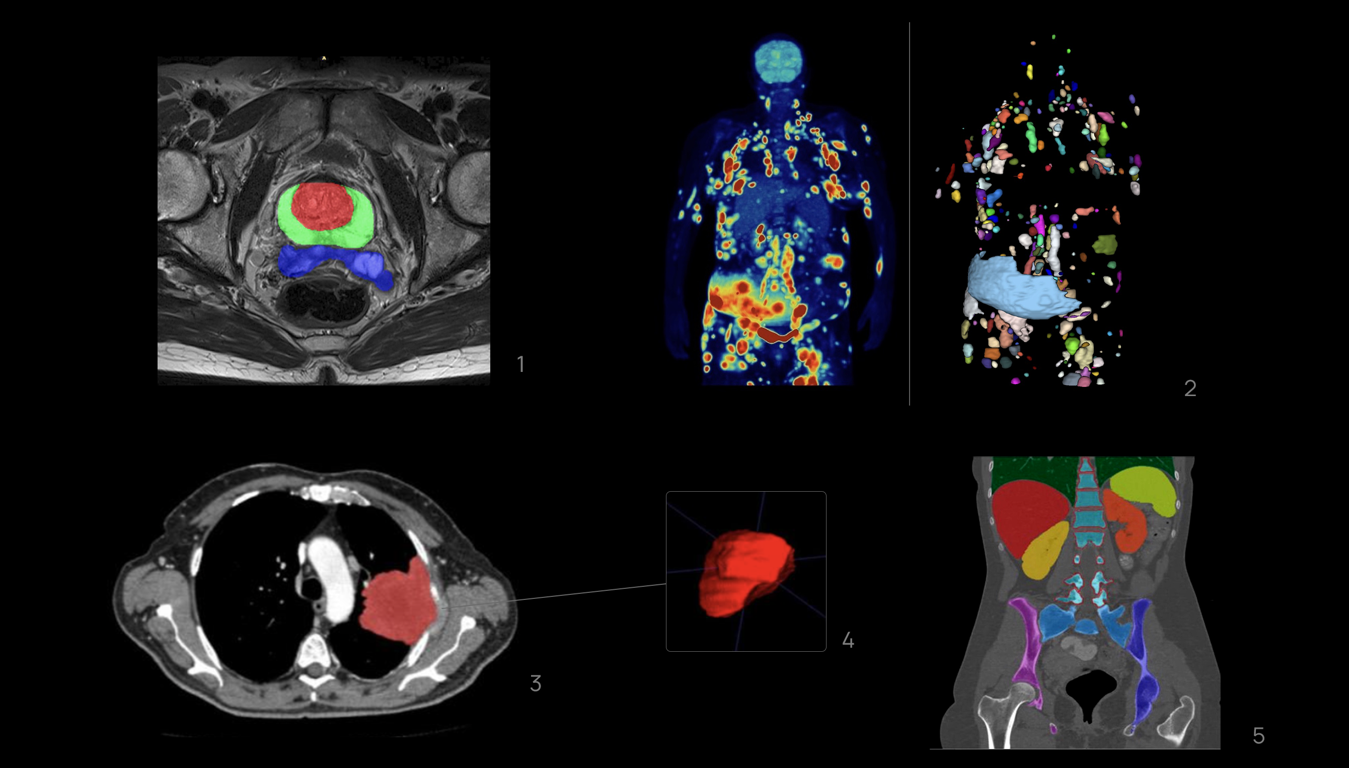
1-Segmentation of the central zone, peripheral zone of the prostate and seminal vesicles on Axial T2-wheited magnetic resonance imaging.
2-Whole body 18-FDG PET/TC imaging exam of a patient with DLBCL in the coronal plane(left) and segmentation mask where every lesion is a unique label (right).
3, 4-Segmentation of the left lung tumor mass on axial Computerized Tomography slices with 3D reconstruction.
5-Segmentation of various anatomical structures in a coronal plane on a Computerized Tomography study.
References
- deSouza NM, van der Lugt A, Deroose CM, et al. Standardised lesion segmentation for imaging biomarker quantitation: a consensus recommendation from ESR and EORTC. Insights Imaging. 2022;13(1):159. Published 2022 Oct 4. doi:10.1186/s13244-022-01287-4
Authors
Raúl Yebana – Image Analysis Manager
Víctor Climent – Image Analysis Technician