ProCAncer-I
Blog
Tags:
CancerProCAncer-I

The ProCAncer-I project celebrated its 5th Consortium Meeting in Turin on 7-8 November 2022, bringing together 20 partners, including centers of reference for the treatment of prostate cancer (PCa), world leaders in artificial intelligence (AI) and innovative SMEs, with recognized expertise in their respective domains.
The objective is to design, develop and sustain a cloud-based, secure European Image Infrastructure with tools and services for data handling. The platform will host the largest collection of PCa multi-parametric (mp)MRI, anonymized image data worldwide (>17,000 cases), based on data donorship, in line with EU legislation (GDPR).

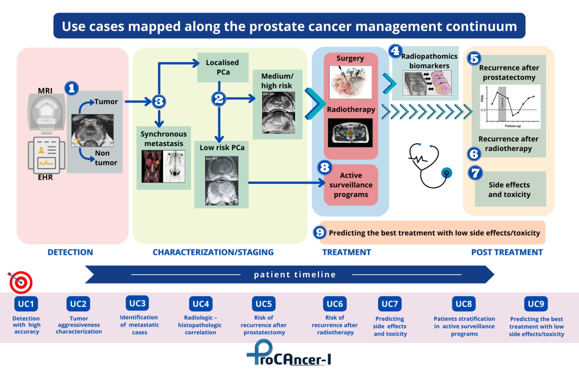
Another main objective of ProCAncer-I is to develop advanced AI models to address unmet clinical needs along the care continuum; i.e. from early diagnosis, to the prediction of metastases and the prediction of response to treatment.
To achieve this, partners will generate a large interoperable repository of health images, and a scalable high-performance computing platform hosting the largest collection of PCa mp MRIs, but also histological and clinical data to be used for the development of robust PCa AI models.
To ensure the rapid clinical implementation of the models developed, the project’s partners will robustly monitor performance, accuracy, and reproducibility in the context of well-designed prospective evaluation studies.
Quibim set out to transform prostate diagnosis and monitoring by developing a new non-invasive imaging tool using MRI data and advanced computer models to investigate the prostate anatomy in extreme detail.
The company has developed AI algorithms for prostate gland segmentation (peripheral zone, central gland, seminal vesicles) with PI-RADS 2.1 parcellation, that can be modified by the experts to store verified annotations and that will be available for the project.
The annotation tool allows to have a concurrent annotation from multiple users, keeping an audit trail that makes secure and reliable use of these data.
In ProCancer-I, Quibim is leading key technical developments, such as the implementation of a monitoring, logging, and retraining system for the development of the AI models, and the application and refinement of deep learning methods for the semi-automated segmentation of images.
Quibim is also the coordinator of the regulatory, exploitation and sustainability activities of the ProCAncer-I platform. These tasks involve exploring the clinical actions regarding the clinical approval of the developed AI models.
Specifically, Quibim is implementing three tools and models at the ProCAncer-I project:
Implementing an image and data annotation tool.
Developing a system for the monitoring, logging, and retraining of AI models.
Building AI models for the automatic segmentation of the prostate.
Over the past few months, Quibim has focused on adapting and integrating the Quibim Precision® annotation environment into the ProCAncer-I platform. In the project, 5% of the cases uploaded to the platform are being annotated through the segmentation of prostate gland and the lesions.
For this purpose, the functionalities of the annotation environment developed by Quibim are being used. This tool includes some manual annotation functionalities, such as the brush tool, which allows precise delineation of the regions of interest.
In addition, for the segmentation of the prostate gland, three different areas are being delineated, including the central/transitional zone, the peripheral zone and the seminal vesicles. Segmenting all these structures from scratch is a time-consuming task. To speed up this segmentation, Quibim has integrated one of the functionalities of its QP-Prostate® product, the algorithm for the automatic prostate segmentation.
This algorithm generates a pre-segmentation that is reviewed by the radiologists of the consortium, who perform its correction for final upload to ProstateNET.
All these annotations are stored in DICOM Seg, a standard format of the DICOM standard which allows storing in a single file a binary map of all the annotations made on a specific series, plus additional metadata, such as the type of annotation (automatic, semi-automatic or manual), the author or the date, together with the reference to the specific series, imaging study and patient. This facilitates the findability, interoperability, and reusability (FAIR principles) of these annotations.
In summa, the novel infrastructure created in ProCAncer-I will help to answer specific clinical questions, supporting precision care through PCa’s continuum.
Website
CORDIS Link:
https://cordis.europa.eu/project/id/952159

This project has received funding from the European Union’s Horizon 2020 research and innovation programme under grant agreement No 952159.