Imaging biomarker panels: setting a new paradigm in oncologic patient care 

Blog
Tags: Biomarkersimaging biomarkersImaging dataMedical imagesPanel reportTreatment

In the evolving and complex landscape of oncology, imaging biomarkers are crucial for tailoring and refining clinical decisions for an individual patient.  Imaging biomarker panels are at the heart of this transformation, converting raw imaging data into insightful predictions. These panels provide insights into treatment response, risk stratification, prognosis, and potential adverse effects.  

They may address pivotal questions such as: Will the patient respond to immunotherapy or targeted treatments?  

The emergence of imaging biomarker panels, with their power to deliver personalized, predictive insights, is an exciting advancement in oncology.


What are biomarkers?

A biomarker, or biological marker, is a measurable indicator that reflects normal biological processes, pathogenic processes, or responses to therapeutic interventions. Although traditionally associated with genetic or molecular traits, biomarkers span a broad spectrum and can be determined using various methods. These include assessments of body fluids such as plasma, serum, or urine to more invasive techniques requiring tumor tissue for comprehensive analyses involving immunohistochemistry, DNA, and RNA.  

Biomarkers serve multiple roles, with prognostic and predictive capabilities being the most significant. Prognostic biomarkers provide insights into a patient’s overall outcome regarding a specific disease, such as cancer. They serve as a valuable guide for clinicians in understanding the likely progression of a patient’s condition. On the other hand, predictive biomarkers offer a view into a patient’s possible response to a specific therapeutic intervention. They aid in tailoring treatment plans to individual patients and reducing the occurrence of adverse effects for optimal results. 

Imaging biomarkers provide a non-invasive method to measure biological processes, furnishing insights into disease progression and treatment effectiveness with consistent correlations to patient outcomes. They have become an integral part of the standard-of-care procedure at every stage of disease management. With a minimal turnaround time, they offer indispensable insights into disease progression and treatment efficacy.


The era of imaging biomarker panels in oncology

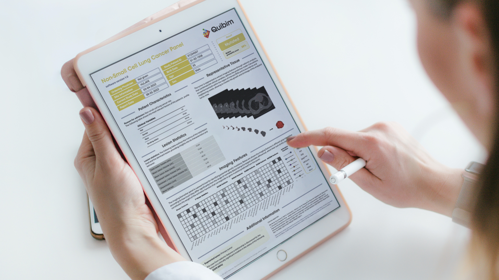
An imaging biomarker panel is a collection of markers identifying various patient subgroups within a disease spectrum. These panels bring complementary information and accuracy to the clinical decision-making process, integrating all data sources into a single report for patient individualization, improving the precision of clinical decision-making.   

Radiomics employs advanced algorithms to extract quantitative features from medical images, revealing patterns often invisible to the human eye. This method can be seen as the imaging counterpart to molecular diagnostics, which interprets molecular signatures to identify diseases. By leveraging routine imaging data from modalities like CT, MRI, and PET, radiomics can isolate unique features that offer insights into the heterogeneity of tissues. These insights often have strong correlations with patient outcomes. Such features might arise from specially designed formulas known as radiomics features or be produced by artificial intelligence (AI) architectures that correlate with clinical endpoints, referred to as imaging biomarkers.  

Imaging biomarkers in clinical trials have become critical, offering a non-invasive way to measure treatment response, stratify patients, and reduce trial risks by improving patient selection for new therapies. Imaging biomarkers also play a significant role in preclinical cancer imaging, helping researchers better understand disease mechanisms and evaluate the effectiveness of novel treatments.


Biomarker panels in cancer imaging: Advancing precision medicine 

In oncology, biomarkers for cancer imaging have reshaped how clinicians assess and treat various cancers. By integrating imaging biomarker panels, oncologists can: 

  • Identify subgroups of patients likely to benefit from specific therapies.
  • Predict responses to treatment, including immunotherapies. 
  • Optimize patient selection in clinical trials, leading to better outcomes.

For instance, research teams like Quibim are pushing the boundaries by developing AI-powered analysis of biomarker panels that improve the reliability of these predictions. Innovation thrives as new imaging biomarkers and AI algorithms are being developed as complementary drug development tools and companion diagnostics and, in the case of Quibim, advancing the approach to a spectrum of diseases from lung, prostate, colorectal, and breast cancer to Alzheimer’s and pediatric cancers to multiple sclerosis.

Inspired by the growing evidence supporting the potential of imaging biomarkers, Quibim proposes an entirely new category of imaging biomarker panels, each customized to specific indications. These panels capture the unique imaging signature of each patient, providing specific and personalized predictions. This approach has pivoted the paradigm, prioritizing individuality and driving precision in clinical decision-making.

MicrosoftTeams-image (55)

Transforming clinical studies with imaging classifiers  

Our primary partner in this endeavor is the biopharma industry. Together, we are creating validated imaging classifiers to identify patients more likely to benefit from targeted therapy. Imaging classifiers can revolutionize clinical trial design by stratifying patients for enrollment in phase 2/3 trials. These patients may benefit from the experimental treatment, maximizing the potential for successful outcomes. Furthermore, creating AI tools to improve patient selection helps to de-risk pivotal trials by identifying those likely to respond to treatment and experience fewer severe adverse effects. 


Overcoming challenges in imaging biomarker validation 

Despite the potential of imaging biomarkers, challenges remain. Many AI models developed are based on single-center studies and, therefore, need external validation. There is a pressing need for large meta-analyses and multi-center studies to generate the evidence required to impact clinical guidelines and redefined treatment indications. Several publications show the potential of radiomic features and imaging biomarkers in predicting clinical outcomes. The emerging paradigm in oncology is the identification and application of radiomic features and imaging biomarkers to predict clinical outcomes.  

Recently, several studies have demonstrated their potential in providing personalized care to patients.  For instance, Cerdá et al. employ a methodology that uses MR quantitative diffusion to distinguish between benign and malignant neuroblastic tumor profiles in children, establishing essential groundwork in pediatric oncology1.

Progressing from this, Rodríguez-Ortega and colleagues utilized multiparametric MR imaging radiomics to predict myometrial invasion in patients with endometrial cancer accurately. Their research underscores the value of merging machine learning with texture features from MR images, challenging the traditional biases in MR reports.  

Bruixola et al. and Marti-Bonmati et al. underscore similar promises in head and neck and pancreatic cancer diagnosis and treatment. Their findings suggest that deep learning techniques hold immense promise in elucidating specific tumor characteristics. Another promising avenue is the exploration of multiple biomarkers, including exosomal mRNA expression, tumor mutational load, and radiomic features, to anticipate responses to immunotherapy in patients with metastatic non-small cell lung cancer (NSCLC), as discussed by Del Re et al. 5 Pushing the frontier even further, Ferrer-Lores et al. explored the integration of genetic alterations with radiomic features in imaging, pinpointing the potential of their amalgamation in predicting first-line treatment responses in DLBCL patients6. 

These pioneering research pieces highlight the game-changing potential of radiomic features and imaging biomarkers, especially when combined with advanced computational methods and genetic data, in revolutionizing oncology by enabling precision diagnosis, prognosis, and patient-specific therapeutic strategies. 


A future of personalized patient care

Integrating imaging biomarkers into an AI-based model is sketching a new trajectory in oncology.  By improving the precision of clinical decision-making, personalizing treatment plans, and facilitating drug development, we are edging closer to a future where each patient receives personalized care tailored to their unique condition. As we refine and validate these tools, we move closer to a future where every patient gets the care they need, when they need it, designed specifically for them. Let’s embrace innovation for medical discovery. The journey has just begun.


References:

  1. Cerdá Alberich L. et al. (2020) A Confidence Habitats Methodology in MR Quantitative Diffusion for the Classification of Neuroblastic Tumors. Cancers (Basel). 2020 Dec 21;12(12):3858. doi:10.3390/cancers12123858.
  2. Rodríguez-Ortega A, et al. (2021) Machine Learning-Based Integration of Prognostic Magnetic Resonance Imaging Biomarkers for Myometrial Invasion Stratification in Endometrial Cancer. J Magn Reson Imaging. ;54(3):987-995. doi:10.1002/jmri.27625
  3. Bruixola G. et al. (2021) Radiomics and radiogenomics in head and neck squamous cell carcinoma: Potential contribution to patient management and challenges. Cancer Treat Rev;99:102263. doi:10.1016/j.ctrv.2021.102263
  4. Marti-Bonmati L., et al (2022) Pancreatic cancer, Radiomics and Artificial Intelligence: A Review. Br J Radiol. Jun 10:20220072. doi: 10.1259/bjr.20220072. Del Re M., et al. (2021). A multiparametric approach to improve the prediction of response to immunotherapy in patients with metastatic NSCLC. Cancer Immunol Immunother. Jun;70(6):1667-1678. doi: 10.1007/s00262-020-02810-6.
  5. Ferrer-Lores B. et al. (2023) Prognostic value of genetic alterations and 18F-FDG PET/CT imaging features in diffuse large B cell lymphoma. Am J Cancer Res. Feb 15;13(2):509-525.