Imaging, AI and radiomics to understand and fight coronavirus Covid-19
Blog
Tags:
BiomarkersCovid-19Medical ImagingRadiomics
There is currently no effective cure for this virus and there is an urgent need to increase global knowledge in its mechanisms of infection, lung parenchyma damage distribution and associated patterns.
Artificial Intelligence and radiomics applied to X-Ray and Computed Tomography are useful tools in the detection and follow-up of the disease.
In December 2019 the city of Wuhan (China) became the center of a pneumonia outbreak of an unknown cause with global implications. In early 2020, Chinese scientists isolated a novel coronavirus (CoV), from patients in Wuhan, formerly known as 2019-nCoV1 and now renamed as Covid-19 by the World Health Organization (WHO). Patients infected with this strain present a wide range of symptoms2, most seem to have mild disease, with about 20% appear to progress to severe disease, including pneumonia, respiratory failure and in around 2% of cases death3. Common signs of infection include respiratory symptoms, shortness of breath and breathing difficulties, fever and cough4.
Coronaviruses (CoV) are a large family of viruses that cause illness ranging from the common cold to more severe diseases such as Middle East Respiratory Syndrome (MERS-CoV) and Severe Acute Respiratory Syndrome (SARS-CoV). This novel coronavirus (nCoV) is a new strain not previously identified in humans. Although this outbreak had its start in China, today there are several countries around the world with identified cases, making it a worldwide public health concern.
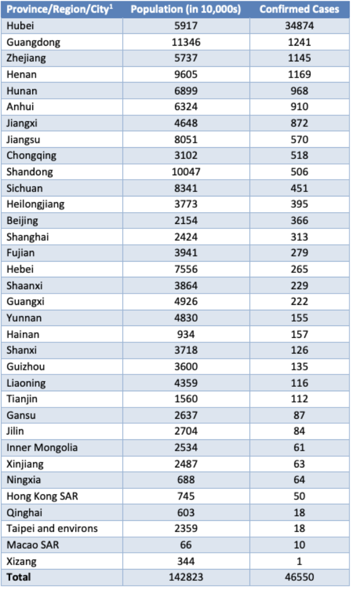
Table 1. Confirmed cases of COVID-19 acute respiratory disease reported by provinces, regions and cities in China, 13 February 2020*
There is currently no effective cure for this virus and there is an urgent need to increase global knowledge in its mechanisms of infection, lung parenchyma damage distribution and associated patterns, not only for disease detection or to complement the diagnosis, but also to support the design of a curative therapy. AI and radiomics applied to X-Ray and Computed Tomography are useful tools in the detection and follow-up of the disease. As stated in5, conspicuous ground grass opacity lesions in the peripheral and posterior lungs on CT images are indicative of Covid-19 pneumonia. Therefore, CT can play an important role in the diagnosis of Covid-19 as an advanced imaging evidence once findings in chest radiographs are indicative of coronavirus. AI algorithms and radiomics features derived from Chest X-rays would be of huge help to undertake massive screening programs that could take place in any country with access to X-ray equipment and aid in the diagnosis of Covid-196.

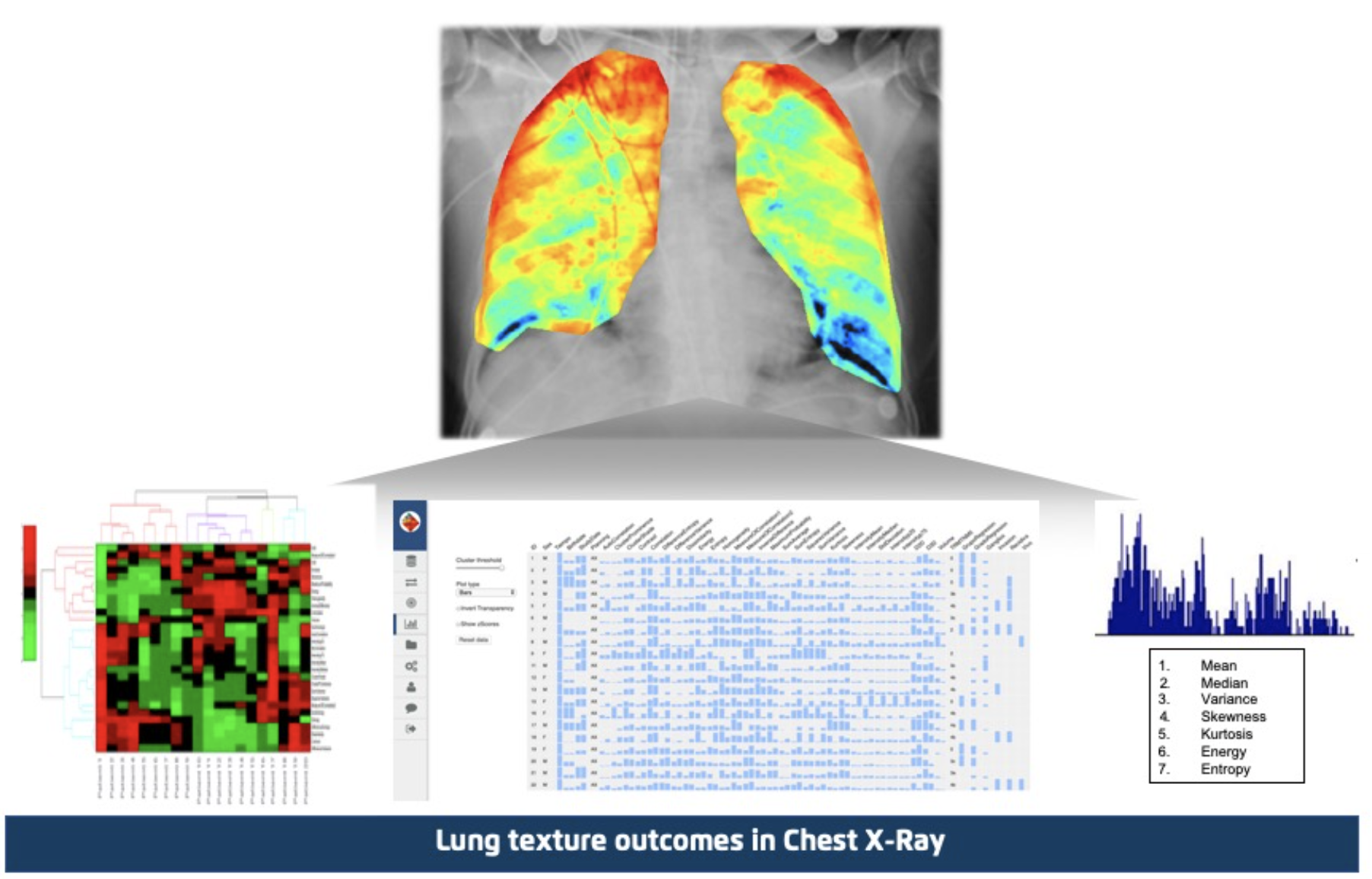
FIGURE 1: Quibim – Quantitative Structured Report – Chest X-Ray Classifier
In order to speed up the discovery of disease mechanisms, Quibim’s Chest X-Ray Classifier (Figure 1) can be used to detect abnormalities and extract textural features of the altered lung parenchima that could be related to specific signatures of the Covid-19 virus. We have combined all our knowledge in AI and radiomics in this novel analysis pipeline specifically designed to extract disease patterns. First, the Chest X-Ray is automatically analyzed using a deep learning classifier to provide an abnormality score between 0 and 1. Any abnormality score above 0.3 is considered as an abnormal case. After this initial analysis, lungs are automatically segmented using a Mask R-CNN like convolutional neural network architecture and finally, a massive extraction of texture features is applied (figure header). This pipeline has been completely automated and will serve to provide additional information to the diagnosis of Covid-19.
Quibim is committed to provide access to our existing AI technology to find new diagnostic tools and ways to understand the mechanisms and aggressiveness of the disease, contributing to the efforts to find a cure. Any clinician can fill this form created by Quibim to get free credentials for the use of the AI Chest X-Ray classification analysis technology available in the Quibim Precision® Cloud platform. This research tool is offered to any doctor worldwide with the need of analyzing Chest X-Rays with suspicion of Covid-19.
References: