Cancer is the first cause of death globally. Cancer occurs when healthy cells become tumor cells, which multiply in uncontrolled growth. These cells can overcome their habitual limits and invade other regions of the body in a process called metastasis, which, in turn, is the first cause of decease due to cancer. DIPCAN is a national project in Spain focused on the digitalization and comprehensive management of personalized medicine in cancer. The main objective of DIPCAN is to categorize the metastatic cancer patient through the development of an artificial intelligence algorithm that helps evolve the understanding, diagnosis and treatment choice of cancer patients. Therefore, this study aims to simplify the decision making during the disease management of each patient, and offers a guide in healthcare policies for good practices. This project is an interdisciplinary study where the coordinated action from partners specialized in different areas of the healthcare sector provides multimodal data combining clinical, phenotypical, pathological, radiomics, and genomics features to predict outcomes that move the patient closer to precision medicine.
The project is intended to collect data from 2000 metastatic patients in a common database where all partners can share their analysis to create a global centralized platform. The current DIPCAN partners are Eurofins Megalab, Fundación MD Anderson Internacional España, Genomcore, Artelnics, Quibim, Pangaea Oncology, and Atrys Health.
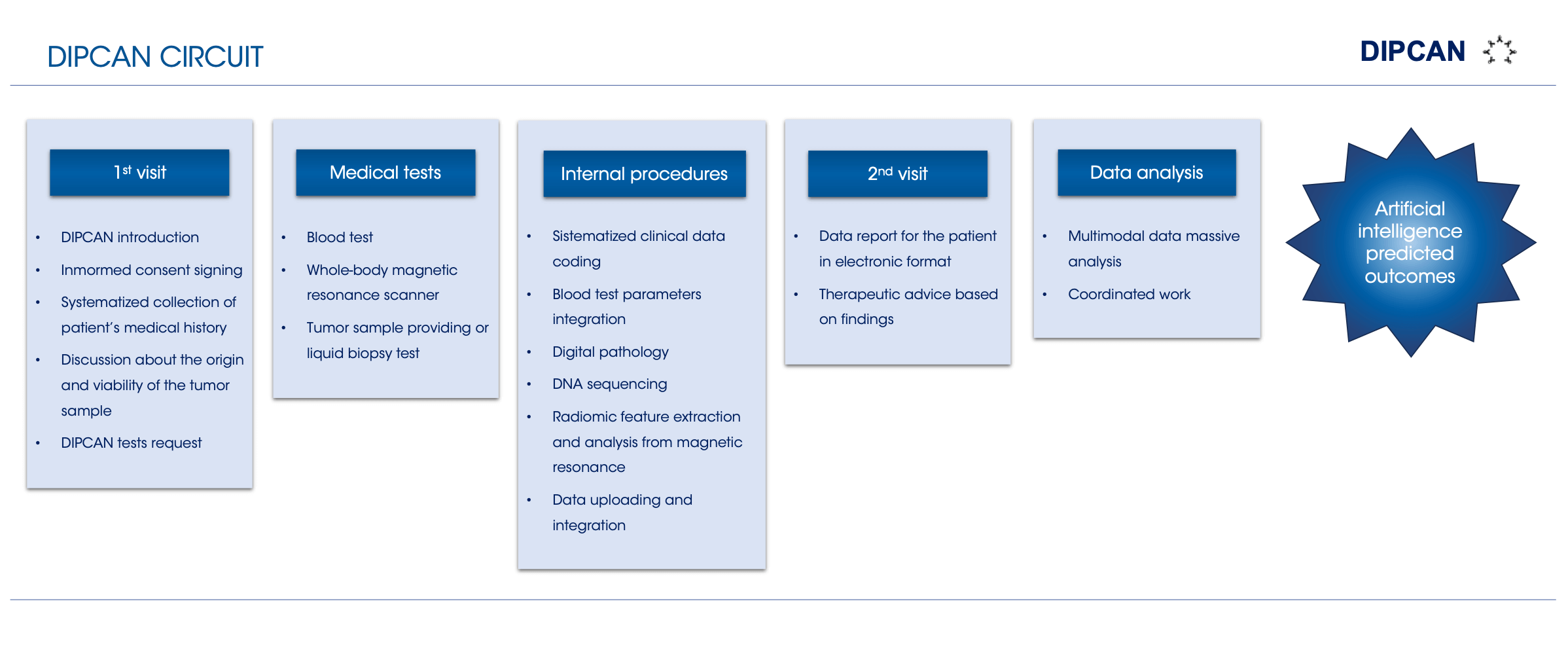
Patient and data circuit
Metastatic cancer patients can join the study through DIPCAN’s website. Once the patient has been registered and has signed the informed consent, they attend the first oncological appointment. The patient provides a primary tumor sample and undergoes several medical tests, including magnetic resonance imaging (MRI), blood test and tumor sample analysis or liquid biopsy. Moreover, collected samples are used for radiomics, genomics and histopathological studies. Finally, the patient will be dated for a second appointment with the oncologist to do a follow-up and consult the results.
Quibim as a key medical imaging partner
Quibim is the reference company in medical imaging in charge of the MRI processing from metastatic cancer patients in DIPCAN. Quibim develops algorithms to extract imaging biomarkers that are invisible to the human eye. These biomarkers are potential prediction points to establish patterns among disease characteristics and patients. This way, predictive models can be implemented for medical purposes.
In this context, the specific objectives of Quibim in DIPCAN are:
For this purpose, Quibim has previously coordinated the implementation of image acquisition protocols adapted to the project requirements for the magnetic resonance scanners in the acquisition centers.
Manual segmentation is a time-consuming task that hinders radiologists’ workflow. Thanks to the use of AI and Deep Learning, this process can be automated. To achieve this goal, Quibim is developing a deep learning model, trained from manual segmentations, to automatically detect and delineate any tumoral lesion in whole-body MRI. Then, radiomics analysis from these segmentations will extract quantitative features, including radiomics and diffusion analysis characteristics. The combination of both algorithms provides a complete automatic tool for tissue characterization.
On the one hand, diffusion MRI represents the flux of water molecules through body tissues, which helps understand specific tissue behaviors, such as tumor tissues. On the other hand, radiomics analysis consists of the extraction of quantitative features intrinsic to the image that the human eye cannot detect. These features are calculated from the MRI lesion segmentations, which delimit the regions of interest (ROIs). Radiomic features provide information about the shape, volume, histogram (intensity value distribution), spatial intensity relationship, and information acquired from statistical methods. Hence, in DIPCAN, all these data are extracted from the malignant masses ROIs and introduced as input to the artificial intelligence predictive model. This way, the biomarkers that characterize the metastatic patient quantitatively can be found and related to gene mutations, phenotypes, or even appropriate treatments for each patient.

Together with the rest of the technical partners, Quibim advises designing an appropriate predictive algorithm to garner the best outcomes for cancer patients.
Thereby, DIPCAN project aims to develop an artificial intelligence tool to prevent, diagnose and ease the treatment choice of each metastatic cancer patient, as well as to contribute to the improvement of the oncology ecosystem.