Advancing liver disease assessment: a breakthrough in automatic liver segments and quantification using Convolutional Neural Networks (CNN)

Blog
Tags: AIHealth TechLiver diseasePrecision Medicine

Liver diseases, such as non-alcoholic fatty liver disease (NAFLD) and non-alcoholic steatohepatitis (NASH), pose significant challenges in diagnosis and monitoring.

Evaluating liver deposits, including fat and iron, is crucial in assessing these chronic diffuse liver diseases. However, existing liver segment methods have limitations in terms of computational costs, execution times, and generalizability to different liver shapes and sizes. This scientific paper introduces a novel methodology utilizing CNN for automatic liver segmentation and quantification in magnetic resonance images (MRI), offering a promising solution to address these challenges.

CNN-based model for liver segmentation and automatic quantification in magnetic resonance imaging (MRI)

The proposed CNN-based model employs an encoder-decoder structure with four convolutional blocks on each branch. Deep supervision and normalization techniques are incorporated to enhance network generalization and boundary detection. The training process involves data augmentation and a 5-fold cross-validation strategy to assess the network’s robustness and generalization to new data.

The performance of the CNN-based liver segmentation model is evaluated using a retrospective multicenter and international dataset of patients with suspected diffuse liver disease. The results demonstrate high accuracy, with a median Dice coefficient of 94% and a false discovery rate of 4%. The CNN-based segmentation also exhibits low relative errors in quantifying proton density fat fraction (PDFF) and R2* relaxation rate, essential biomarkers for the diagnosis and treatment monitoring of liver diseases.

The study further examines the heterogeneity of fat and iron distribution in the liver and confirms the model’s ability to generalize to cases with various distributions. External validation using an independent dataset from different centers and scanners demonstrates the reproducibility of the model across various imaging settings.

Improving diagnosis through a reliable and efficient solution for liver and fat quantification in MR images

This automated segmentation approach can improve the diagnosis and monitoring of chronic diffuse liver diseases, providing additional information to radiologists and reducing the limitations associated with manual segmentation methods.

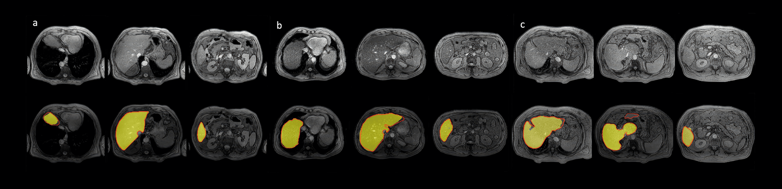
The breakthrough methodology presented in this scientific paper demonstrates the effectiveness and generalizability of a CNN-based model for automatic liver segmentation and quantification. By accurately capturing the liver parenchyma, this approach enables precise assessment of fat and iron deposits, essential for diagnosing and monitoring chronic diffuse liver diseases.

The CNN-based model offers a fast and automatic procedure for MR virtual biopsy, significantly enhancing the clinical evaluation of liver diseases and opening new avenues for research and treatment advancements in this field.

Liver segments: Figure Image Advancing liver disease
Figure 1. Segmentations were obtained on three different test cases at different anatomical levels. In yellow the CNN predicted segmentation mask is shown and in red the contour of the ground truth is represented. a Results in a study from one of the centers used for the model development. b Results in a study from the other center used for the model development. c Performance on a study which belongs to a patient with partial hepatectomy

Referece

Precise whole liver automatic segmentation and quantification of PDFF and R2* on MR images. Jimenez-Pastor, A., Alberich-Bayarri, A., Lopez-Gonzalez, R. et al. Eur Radiol 31, 7876–7887 (2021) 

Read more