Advanced imaging biomarkers in prostate cancer – A leap towards precision in risk stratification and BCR prediction
Prostate cancer (PCa) is a prominent health concern, the second most common cancer among men globally, and the second leading cause of cancer death in men1. The complexity and variability in its progression necessitate advanced methodologies for accurate risk stratification and prediction of biochemical recurrence (BCR)2. Numerous risk stratification systems have been devised to assist in differentiating aggressive tumors from indolent ones to mitigate this variability.
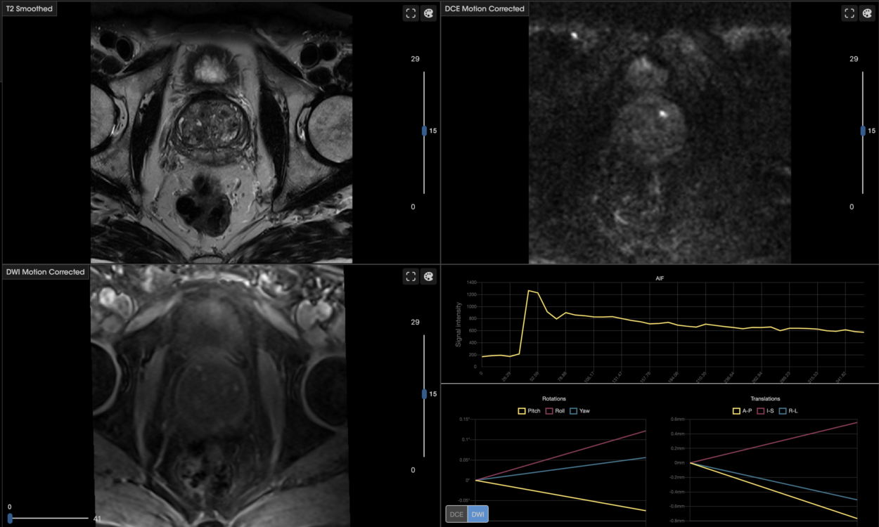
A recent study published in Cancers delves into the potential of utilizing imaging biomarker profiles extracted from magnetic resonance imaging (MRI) to enhance the precision in identifying PCa patients with a higher risk of BCR and to guide treatment decisions more effectively.
A closer look at imaging biomarker profiles
The study aimed to discern imaging biomarker profiles, specifically those involving perfusion/diffusion and radiomic features from MRIs, which could distinguish patients based on their risk and the occurrence of BCR a decade post-diagnosis. The research encompassed 128 patients, with a mean age of 71 years, who were retrospectively evaluated. These patients, diagnosed with localized PCa, underwent neoadjuvant androgen deprivation therapy and radiotherapy.
The primary objectives were to pinpoint radiomic profiles in MRIs taken post-ADT initiation to (1) classify patients with a more severe prognosis based on their risk stratification groups and (2) forecast the occurrence of BCR a decade post-diagnosis. Another exploratory goal was to discern radiomic profiles in patients categorized as high/unfavorable-IR for BCR prediction.
Analyses were conducted on the entire prostate and specific prostate regions to determine if regional analysis would impact the results. The extracted imaging features from MRIs were analyzed for each prostate region and for the whole gland, employing univariate and multivariate analyses. The results indicated that prostate region-wise imaging biomarker profiles, predominantly composed of radiomic features, could discriminate risk groups, and identify patients experiencing BCR. Notably, heterogeneity-related radiomic features were elevated in patients with a worse prognosis and in those experiencing BCR.
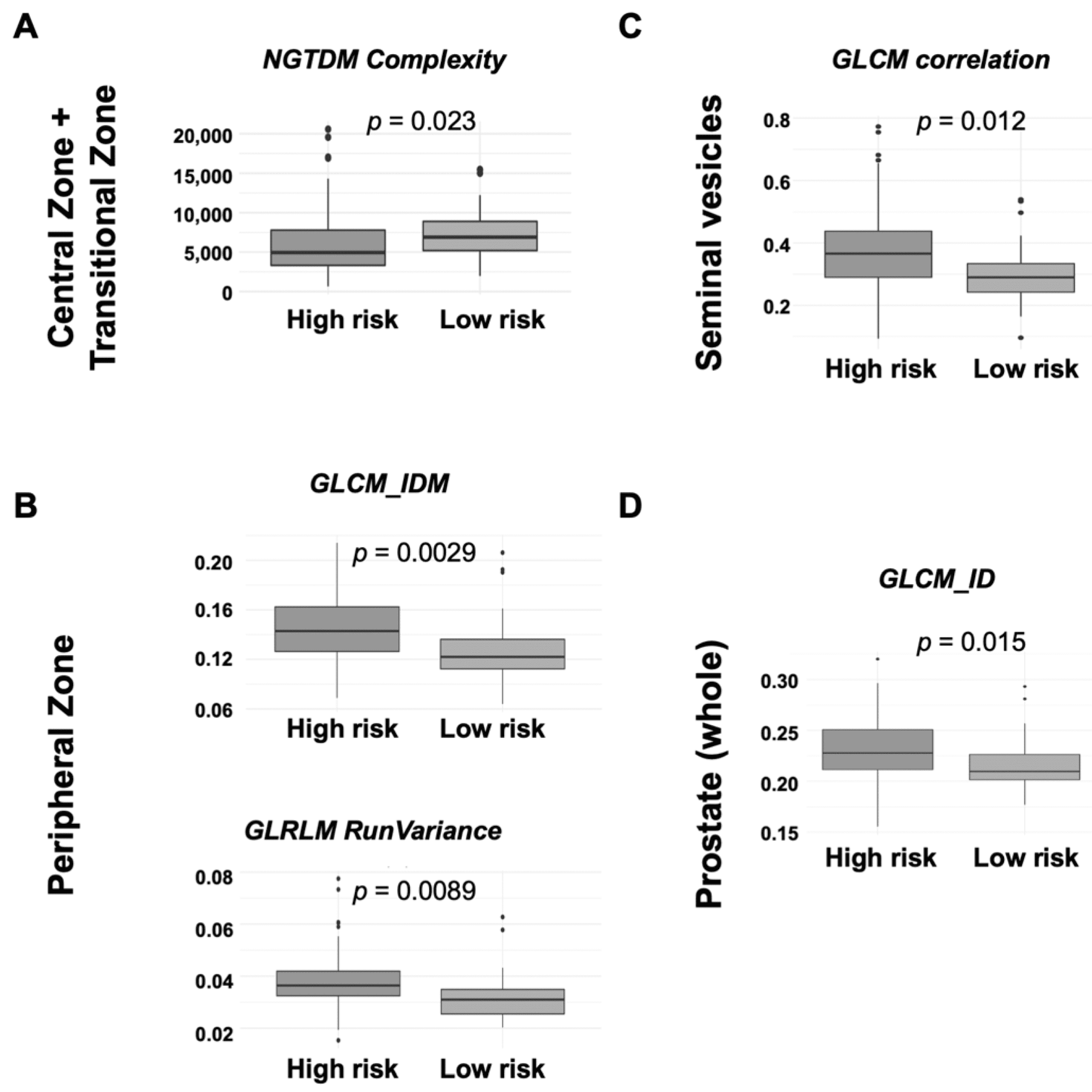
Box plots for imaging (radiomics) biomarkers in each prostate region for risk stratification (arbitrary units). (A) Central Zone + Transitional Zone; (B) Peripheral Zone; (C) Seminal vesicles; (D) Prostate (whole). GLCM = Gray-Level Co-Occurrence Matrix; GLRLM = Gray Level Run Length Matrix; ID = inverse difference; IDM = inverse different moment.
Key findings: radiomic features and predictive ability
The study revealed that imaging biomarkers maintained a commendable predictive ability, with area under the curve (AUC) values exceeding 0.725 in most of the cases. This predictive capability was generally enhanced when clinical data were incorporated, especially for BCR prediction, where AUC values fluctuated between 0.841 and 0.877 for combined models and sensitivity values surpassed 0.960. The models built per prostate region versus the whole gland showed noticeable improvement.
A beacon of hope in PCa management
Radiomics, which enables the extraction of quantitative features from medical images, has garnered attention in recent years, particularly in PCa management. The extracted radiomic features from MRI have proven instrumental in PCa detection, localization, and clinical outcome prediction3-8. The study under discussion further substantiates the potential of radiomics in identifying and localizing prostate cancer on MRI, discriminating between cancer and normal prostate tissue, and associating textural features with BCR following PCa radiotherapy.
Implications and future directions
The study’s findings underscore the pivotal role of prostate region-aware imaging profiles in identifying patients with a worse prognosis and a higher risk of BCR. When amalgamated with clinical variables, these profiles retain superior predictive values, thereby opening avenues for more personalized and effective treatment strategies for PCa patients.
The advancements in imaging biomarker profiles open a future where risk stratification and BCR prediction in PCa patients can be significantly more accurate and tailored. This holds the promise of enhancing therapeutic outcomes and minimizes the risk of over-treatment or under-treatment, ensuring that patients receive the most optimal care based on their individual risk and prognosis.
Final remarks
Distinct imaging biomarker profiles, primarily formulated of radiomic features and tailored to specific prostate regions, demonstrated the capability to categorize patients based on their risk and the emergence of BCR a decade post-diagnosis.
Moreover, these imaging profiles, whether standalone or amalgamated with clinical variables, facilitated the creation of models to prognosticate patient risk and BCR, applicable to both the general population and particularly to patients with high/unfavorable Intermediate-Risk (IR).
As we forge ahead, further research and clinical trials will be instrumental in refining and integrating these methodologies into standard clinical practice, thereby elevating the standard of care provided to PCa patients.
—
Sánchez Iglesias Á, Morillo Macías V, Picó Peris A, Fuster-Matanzo A, Nogué Infante A, Muelas Soria R, Bellvís Bataller F, Domingo Pomar M, Casillas Meléndez C, Yébana Huertas R, et al. Prostate Region-Wise Imaging Biomarker Profiles for Risk Stratification and Biochemical Recurrence Prediction. Cancers. 2023; 15(16):4163.
https://doi.org/10.3390/cancers15164163
References
-
Bergengren, O.; Pekala, K.R.; Matsoukas, K.; Fainberg, J.; Mungovan, S.F.; Bratt, O.; Bray, F.; Brawley, O.; Luckenbaugh, A.N.; Mucci, L.; et al. 2022 Update on Prostate Cancer Epidemiology and Risk Factors—A Systematic Review. Eur. Urol. 2023, 84, 191–206.
-
Mottet, N.; van den Bergh, R.C.N.; Briers, E.; Van den Broeck, T.; Cumberbatch, M.G.; De Santis, M.; Fanti, S.; Fossati, N.; Gandaglia, G.; Gillessen, S.; et al. EAU-EANM-ESTRO-ESUR-SIOG Guidelines on Prostate Cancer-2020 Update. Part 1: Screening, Diagnosis, and Local Treatment with Curative Intent. Eur. Urol. 2021, 79, 243–262
-
Fehr, D.; Veeraraghavan, H.; Wibmer, A.; Gondo, T.; Matsumoto, K.; Vargas, H.A.; Sala, E.; Hricak, H.; Deasy, J.O. Automatic classification of prostate cancer Gleason scores from multiparametric magnetic resonance images. Proc. Natl. Acad. Sci. USA 2015, 112, E6265–E6273.
-
Ginsburg, S.B.; Viswanath, S.E.; Bloch, B.N.; Rofsky, N.M.; Genega, E.M.; Lenkinski, R.E.; Madabhushi, A. Novel PCA-VIP scheme for ranking MRI protocols and identifying computer-extracted MRI measurements associated with central gland and peripheral zone prostate tumors. J. Magn. Reson. Imaging 2015, 41, 1383–1393.
-
Lemaître, G.; Martí, R.; Freixenet, J.; Vilanova, J.C.; Walker, P.M.; Meriaudeau, F. Computer-Aided Detection and diagnosis for prostate cancer based on mono and multi-parametric MRI: A review. Comput. Biol. Med. 2015, 60, 8–31.