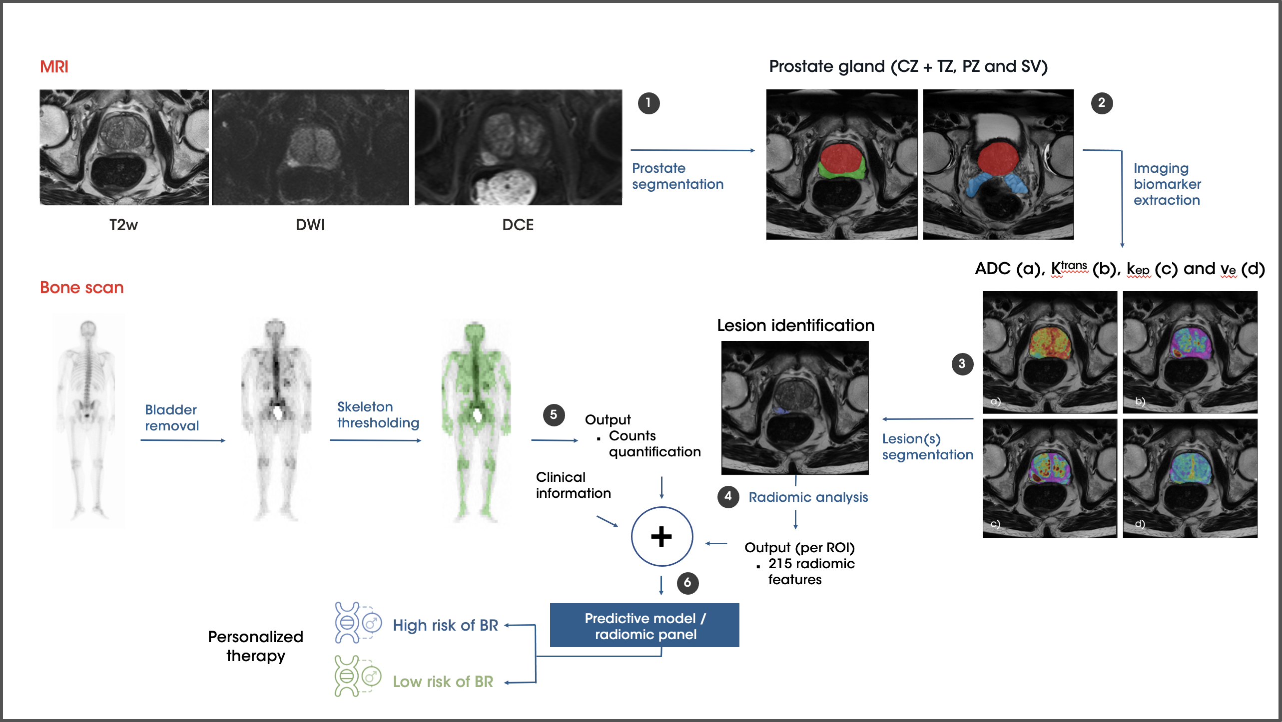
Prediction of metastatic relapse in intermediate/high risk localized prostate cancer patients from staging medical images and clinical variables

Predicting metastatic relapse in prostate cancer through AI.
Application Oncology

The challenge

Prostate cancer (PCa) is the second most common cancer in men worldwide and the second leading cause of cancer death in men. Most localized PCa represents an indolent disease, however, intermediate- and high-risk patients are more likely to die from prostate cancer and its prognosis is more difficult to predict. For these patients, prognosis remains complex, notably as biochemical recurrence (BCR) marks a significant step towards disease progression, raising the risk of metastases. Therefore, accurate risk stratification and swift diagnosis are imperative to enhance treatment decisions and decrease progression incidences.

The solution

This retrospective, multicenter study aims to develop an AI model predicting metastatic relapse in 300 intermediate or high-risk localized PCa patients. It will utilize diagnostic medical images, including multiparametric MRI (T2-W, DWI, DCE sequences), staging bone scans, and clinical data, all consolidated on the QP-Discovery® Platform. Automatic segmentation of prostate gland MRIs and lesion(s) detection followed by the extraction of imaging biomarkers will be performed, supplemented by counts quantification from staging bone scans. In conjunction with clinical variables, this data will inform the predictive model, forecasting patient metastatic relapse.

The outcome

The project aims to predict the likelihood of metastatic recurrence at diagnosis automatically, non-invasively, and in real-time, allowing for a complete patient stratification that would allow tailoring of specific aspects of adjuvant therapy as well as an intensification of initial treatment for localized PCa patients diagnosed as intermediate/high risk.   

Image 01 predict metastasis prostate cancer


This diagram illustrates the methods applied to the collected medical images

  1. We automatically segment the prostate gland’s morphological MR image (T2w) into three key regions: CZ + TZ, PZ, and SV.
  2. We extracted imaging biomarkers from DWI and/or DCE images, depending on the protocol used (bi-parametric or multi-parametric).
  3. Using these biomarkers, we generate probability maps to identify potential lesions, creating a corresponding nosological map.
  4. Pre-segmented lesions are then isolated, and quantitative imaging features are extracted.
  5. In bone scan analysis, we remove the bladder, threshold the skeleton, and quantify percentile counts, focusing on the 92nd percentile as a key biomarker.
  6. Finally, we develop a predictive model using clinical data and extracted imaging features. The model’s output stratifies patients into high or low-metastasis-risk groups.